Conquering coag testing, from basics on up

| Conquering coag testing, from basics on up |
|
|
|
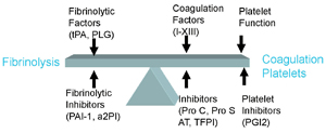
January 2011 William Check, PhD While pathologists are understandably excited about new molecular methods, as evidenced by heavy attendance at molecular sessions last fall at the annual meetings of the ASCP and CAP, at the same meetings it was clear they continue to focus on the basics as well. Coagulation is an example. At both meetings, courses on basic coagulation were heavily subscribed. Interest in coagulation testing stems from its importance and its complexity. “My course was bread and butter hemostasis,” Dorothy M. (Adcock) Funk, MD, medical/laboratory director of Esoterix Coagulation, told CAP TODAY. Dr. Funk presented “An Overview of Coagulation” at the ASCP meeting. “Many pathologists feel they don’t quite understand the complexities of coagulation testing and interpretation. It seems confusing to them. They want to get a better handle on it,” she says, noting that coagulation specialists are “few and far between across the country” yet basic coagulation testing has to be performed in hospital labs. John D. Olson, MD, PhD, one of the speakers at the CAP course, “An Algorithmic Approach to Hemostasis Testing,” expresses a similar view. “For most clinicians and laboratorians these are very commonly ordered tests. People use them all the time. And yet they probably don’t understand the details as well as they could.” Dr. Olson is vice chair and professor of pathology and director of clinical laboratories at the University of Texas Health Science Center at San Antonio. He and his co-presenter, Kandice Kottke-Marchant, MD, PhD, chair of the Pathology and Laboratory Medicine Institute and section head of hemostasis and thrombosis at the Cleveland Clinic, told CAP TODAY that the course was aimed at general pathologists who also cover clinical laboratory functions. “We give them a foundation for running a hemostasis lab,” she says. As a metaphor for the coagulation system, Dr. Marchant displayed an image of Janus, the Roman god of ending and beginning, whose two faces look in opposite directions. “Bleeding and clotting are two faces of the same system,” Dr. Marchant says. “All of hemostasis is checks and balances. Depending on whether pro- or anticoagulant factors predominate, you can have bleeding or thrombosis.” In her introductory talk, Dr. Marchant listed four reasons to order a hemostasis test: to ascertain causes of bleeding, to evaluate the etiology and clinical significance of an abnormal coagulation test result, to monitor the effect of anticoagulants, and to evaluate risk factors for thrombophilia. “One of the crucial things about hemostasis testing is that it can be ordered for many different reasons by many different practitioners,” she says.
It may not always be clear to the lab why a test was ordered. The most commonly ordered coagulation test is the APTT, which is used to screen patients for bleeding problems before surgery or to monitor patients on anticoagulant therapy, such as heparin. “The way you evaluate APTT results differs greatly in these two contexts,” Dr. Marchant says. An elevated APTT in the presurgical patient can trigger further testing, while in a patient on heparin it is the desired outcome. Dr. Marchant reviewed the technical considerations affecting APTT results, all of which are in a CAP monograph (Krishnan J. Sample collection and processing for coagulation. In: Kottke-Marchant K, ed. An Algorithmic Approach to Hemostasis Testing. CAP Press: 2008). CLSI guidelines H21-A5, C28-A2, and H57-A also contain useful information about coagulation testing. One crucial point: The choice of APTT reagent affects the heparin therapeutic range. Dr. Marchant showed published data for two reagents: For one the APTT therapeutic range was 60–80 seconds; for the other it was 100–160 seconds (Brill-Edwards P, et al. Ann Intern Med. 1993;119:104–109). In her course, Dr. Funk too discussed reasons for ordering APTT and prothrombin time: preoperative hemostasis screening; evaluation of the bleeding patient; and monitoring anticoagulant therapy. In addition, she presented extensive information on plasma mixing studies and gave an update on lupus anticoagulants. For preoperative screening, Dr. Funk said, “The best means to determine hemorrhagic risk is an adequate history and physical examination.” A positive medical history, which includes a family history, is 12.5 times more likely to predict hemorrhage than a battery of lab tests. Obtaining a patient’s medication history, including over-the- counter agents and herbal preparations, is also imperative. Many drugs affect hemostasis. Aspirin and nonsteroidal agents are well-known examples. Less well known are SSRIs, certain antibiotics (moxalactam, tobramycin), anticonvulsants, and chemotherapeutic agents. Herbal products and supplements can also alter hemostasis—feverfew, ginseng, Dong Quai, and fish oils among them. In a patient without a bleeding history, the preoperative screen includes APTT, PT, and platelet count. “But these don’t have great value in predicting who will bleed with surgery,” Dr. Funk reiterated. In fact, in Dr. Funk’s recommendations for pre-op hemostasis screening, a person with no history of bleeding who has undergone hemostatic stress should not be screened. In the absence of a history of bleeding with no previous challenge or for a procedure that is not high bleeding risk, a screen may or may not be done. Only when there is a suspicion or history of a bleeding disorder, or if the patient has never been challenged and the procedure has significant bleeding potential, is screening recommended. To evaluate a patient with a history of bleeding, the characteristics of the bleeding episode(s) should be examined—spontaneous or provoked, from single or multiple sites, recent onset or lifelong. “With bleeding from a single site, you always have to think about an anatomical cause,” Dr. Funk says. “In a post-op patient it may be surgically related, such as faulty or missing sutures. If bleeding is related to sutures, we say the patient is deficient in factor XIV (aka suture).” A disorder of primary hemostasis is characterized by “mucosal bleeding”—immediate bleeding, menorrhagia or postpartum hemorrhage, gingival bleeding, or petechiae. This type of bleeding might be due to a platelet defect or to von Willebrand disease. A disorder of secondary hemostasis, which manifests as deep ecchymoses, joint hemorrhage, hematomas, or delayed bleeding, typically results from a factor deficiency. A hemostatic screen to evaluate a bleeding disorder is similar to a pre-op screen—APTT, PT, and platelet count. Additional tests include thrombin clotting time (TCT or TT) or fibrinogen activity and some form of platelet function test, especially for a patient who presents with mucosal bleeding. For a primary bleeding disorder, von Willebrand factor assays are measured as well. Dr. Funk called platelet function testing “problematic,” saying, “It is only sparsely available” and compounding this is the problem that “platelets don’t travel.” Dr. Funk’s lab performs the test but the patient must travel to her laboratory to get his or her blood drawn. In this circumstance, the patient is typically worked up after the bleeding is controlled and after she or he is released from the hospital. Many have a long history of bleeding. “My last patient had been bleeding intermittently for seven years either spontaneously or after surgery,” Dr. Funk told CAP TODAY. The basics—APTT and PT—are what the laboratory can do best. Many conditions can cause an isolated elevated APTT, such as a lupus anticoagulant, deficiency or specific inhibitor of factors VIII, IX, or XI, or heparin therapy or contamination. Dr. Funk noted that lupus anticoagulants rarely cause bleeding. To distinguish a factor deficiency from a specific inhibitor, mixing studies are the best technique. In this procedure, one part patient plasma (unknown amount of factor) is mixed with one part normal plasma (about 100 percent factor level). Correction indicates a factor deficiency. No or partial correction indicates a factor inhibitor. “Mixing studies sound easy, but they must be performed and interpreted correctly or you’ll go down the wrong pathway,” Dr. Funk emphasized. “It is not unreasonable for a routine lab to not perform this test but rather send it out.” Mixing studies can be difficult to perform properly and interpret because they have fairly significant technical requirements for such a “simple” test and neither the method nor the means to interpret results is standardized. Another complication is that some inhibitors, such as lupus anticoagulants, can occasionally show correction. To confuse things further, inhibition can be time dependent—it can appear with time, since some inhibitors, particularly specific factor VIII inhibitors, require incubation for effect. An incubated mixing study (60 to 120 minutes at 37°C) should be considered whenever correction is seen. For a minimally prolonged APTT or PT, mixing studies are even more difficult to interpret and for this reason are not indicated. When a factor inhibitor is suspected through an APTT mixing study, factor VIII (the most common factor associated with a specific inhibitor), IX, and XI assays should be performed at multiple dilutions (after heparin interference has been excluded) and a lupus anticoagulant ruled out as well. An isolated elevated APTT can be due to hereditary factor deficiencies, such as factor VIII (hemophilia A and autosomal vWD), factor IX (hemophilia B), and factor XI (hemophilia C), all of which may be clinically significant. Deficiencies of factor XII, prekallikrein, and high-molecular-weight kininogen, while elevating the APTT, do not carry an increased risk for bleeding. When interpreting the result of the APTT, Dr. Funk reminded participants that a normal APTT may not always rule out mild deficiencies of factors VIII, IX, and XI (factor levels in the 10 percent to 40 percent range). APTT reagents differ in their responsiveness to factor deficiency, similar to how APTT reagents vary in their responsiveness to heparin. Accordingly, laboratorians should determine their reagents’ responsiveness to each factor or ask the reagent manufacturer for this information. The third component of the hemostasis screen—thrombin clotting time or a fibrinogen assay—can be elevated by low or inactive fibrinogen or by anticoagulant drugs such as heparin or direct thrombin inhibitors. Dr. Funk provided several cases to illustrate these principles. One was a 68-year-old man with chronic lymphocytic leukemia referred by his dentist because of gingival bleeding of recent onset. His APTT was elevated; it normalized with a plasma mixing study. Factor VIII was found to be low. “Low factor VIII is one of the most common things you will investigate in the coagulation lab,” Dr. Funk said. Her next test was von Willebrand factor antigen and activity. vWF activity can be measured in two ways, by collagen binding and by ristocetin-induced agglutination of platelets. “Both of these assays have value as they measure different aspects of vWF function, and in our lab we use them both,” Dr. Funk said. In this case vWF antigen and activity were both low. “vWF antigen levels should correlate with factor VIII activity,” Dr. Funk said, since vWF stabilizes factor VIII in the blood. In this case, with new onset bleeding and no family history, the diagnosis was acquired vWD, which is often associated with hematologic disorders. Its lab manifestations are similar to those of hereditary vWD; the distinction is made on clinical grounds. When vWD is suspected at the outset, APTT should not be used as a screen because it can be normal. Another case—a 27-year-old woman who had a pulmonary embolus—illustrated the clinical impact of antiphospholipid antibodies, which Dr. Funk said are the most common cause of acquired thrombophilia and a possible cause of both arterial and venous thrombosis. Standard methods are in place for defining these antibodies. When antiphospholipid antibodies are suspected, the lab should test for a lupus anticoagulant, anti-cardiolipin antibodies, and—the newest test—anti-beta2GP1 antibodies. Establishing persistence is most important in identifying an antiphospholipid antibody as clinically significant; it requires repeating a positive test at 12 weeks. When testing for lupus anticoagulants, Dr. Funk cautioned, no single test is 100 percent sensitive and specific; a panel of assays is frequently necessary. Furthermore, concordance between clot-based and ELISA-based antiphospholipid antibody assays is only about 60 percent; therefore, both must be performed before antiphospholipid antibodies are ruled out. Another caution: It can sometimes be difficult to distinguish lupus anticoagulant from specific factor inhibitors, especially an inhibitor to factor VIII. One way to help resolve this is to perform a factor VIII chromogenic assay, which is not affected by lupus anticoagulant. New laboratory criteria have been published for the detection of lupus anticoagulants (Pengo V, et al. J Thromb Haemost. 2009;7:1737–1740). “Some aspects of these new criteria are controversial,” Dr. Funk noted, such as recommending a 1:1 dilution of patient and pooled normal plasma when testing is performed in patients with an INR between 1.5 and 3.0. Dr. Funk next turned to fibrinolysis, which she called “the mirror image of hemostasis.” In coagulation a clot is formed; in fibrinolysis the clot is broken down. Just as disorders of fibrin clot formation can lead to either bleeding or thrombosis, so can disorders of fibrinolysis. In particular, bleeding can result from deficiencies of alpha-2-antitrypsin or PAI-1. Fibrin clots are easily lysed when there is an extreme lack of factor XIII activity, due either to a deficiency or an inhibitor. Dr. Funk warned against screening for factor XIII deficiency using the urea-solubility assay, which she said is not useful as it will only be abnormal when factor XIII levels fall below about two percent. “Perform a factor XIII activity assay,” she advises. “Simple tests with difficult problems” is how Dr. Olson describes the prothrombin time and activated partial thromboplastin time assays. Both have long been in common use—the PT since 1935 and the APTT since the early 1950s. Even so, Dr. Olson said, “When you look closely at these tests and how they are used, there are places where people can get confused about applications. The devil is in the details.” Possible causes of an elevated APTT include factor deficiency, nonspecific inhibitors such as lupus anticoagulant, specific factor inhibitors, and medication (heparin, direct thrombin inhibitors). Dr. Olson shared an algorithm for evaluating an abnormal APTT, saying, “Every cause requires a different action on the part of the clinician” and laboratory results contribute greatly to the clinician’s decision. A mildly prolonged APTT does require action, Dr. Olson says. “In most labs now, if the APTT is prolonged five seconds or less, we don’t even do a mixing study. It is more likely to be confusing than helpful. However, evaluation is necessary via a different approach.” The PT can be used to evaluate a potential inherited bleeding condition. However, Dr. Olson said, “An inherited bleeding diathesis is a rare event. Most bleeding disorders are acquired.” Further, the three most common inherited bleeding disorders—factor VIII and factor IX deficiency and vWD—are not detected by the PT. Since vWD affects only factor VIII, it prolongs the APTT but not the PT. “Liver disease and consumptive coagulopathies are probably the most common acquired disorders detected by the prothrombin time,” Dr. Olson said. “Multiple factors are low in acquired disorders.” With regard to mixing studies, Dr. Olson finds that most people associate time dependence with an inhibitor to factor VIII. However, he said, the clinical setting is important. “If you are taking care of patients with hemophilia, among those who have factor VIII inhibitors we will find that the majority, as high as 85 to 90 percent, demonstrate time dependence. So it’s true that in patients with hemophilia it’s common to find time-dependent factor VIII inhibitors.” He continued: “However, if we look at it from the lab side, when we see a prolonged APTT and a mixing study that shows a time-dependent inhibitor, the inhibitor is far more likely to be lupus anticoagulant. Even though only about 10 percent of lupus anticoagulants are time dependent, the presence of lupus anticoagulant in the population is so high that it’s far more likely that time-dependent inhibition will be due to lupus anticoagulant.” Dr. Olson has calculated that time-dependent inhibition in an unselected population is about 160 times more likely to be due to lupus anticoagulant than to a specific inhibitor of factor VIII.
Dr. Olson also advocates standardizing APTT reagents. “In our lab, not in all labs, when we evaluate and choose a reagent, we like to know that the reference interval doesn’t deviate greatly from what we’ve been using, that the sensitivity to deficiencies of factors VIII and IX is acceptable—in the 25 to 30 percent range—and that sensitivity to heparin is similar to our prior reagent. Once we have chosen a reagent, we check its sensitivity to other factors and to lupus anticoagulant.” It is also important to appreciate the correlation—or rather, noncorrelation—between the APTT used to detect heparin and a heparin assay, such as the factor Xa inhibition assay. (“I am on a campaign to get people to talk about this as a heparin assay,” Dr. Olson says, “and not talk about the method for quantifying heparin. Anti-Xa is our method to detect the concentration of heparin.”) Patients within the heparin therapeutic interval can have a twofold (or more) range of APTT times. “The two tests measure completely different things,” Dr. Olson says. “So there is no nice correlation. The reason is that patients and methods vary widely in their response to heparin.” The lack of correlation between these two methods is a conundrum. “The number of labs reporting that they measure heparin is growing,” Dr. Olson says. “But this does not mean they are using a heparin assay for routinely monitoring unfractionated heparin therapy, although it is also true that there are more labs using a heparin assay. Many labs monitor heparin with APTT but have a heparin assay available for when the APTT is unacceptable.” A problem arises because, unlike other assays where introducing a new assay results in dropping the old one, introducing a heparin assay does not result in eliminating the APTT. “Some clinicians still order the APTT for monitoring heparin,” Dr. Olson says. “But because there is such poor correlation between the heparin assay and the APTT, there will be times when the heparin assay looks like it is within the therapeutic interval and the APTT could be outside the therapeutic interval.” Then the clinician has a dilemma: whether to change dose or not. “When clinicians call me about this, my advice is to stop using the APTT for measuring heparin,” Dr. Olson says. “It just causes confusion.” In Dr. Funk’s course, the last topic she covered was thrombophilia, defined as a disorder associated with an increased tendency to develop (venous) thromboembolism. Since thrombophilia is a “tendency,” there are many people with thrombophilia who will never develop a thrombosis. Dr. Funk underlined the impact of thrombophilia when it manifests clinically. She cited venous thrombosis “as a common disorder in the population, with an incidence of one or two cases per 1,000 persons.” Deep vein thrombosis, or DVT, affects more than 2 million Americans per year. Pulmonary embolus causes 10 percent of hospital deaths; it is the most common cause of preventable hospital death. Genetic risk factors predisposing to thrombosis include deficiencies of antithrombin, protein C, or protein S, or the presence of the following specific mutations: prothrombin 20210 or factor V Leiden. “Antithrombin (AT) deficiency is the most important genetic risk factor for thrombophilia,” Dr. Funk said. It carries a 24-fold increased risk. Heterozygous factor V Leiden has a lower risk potential (three- to eightfold increase), but it is more common in the population. Acquired risk factors for thrombophilia include antiphospholipid antibodies, cancer, surgery, estrogens (oral contraception, hormonal replacement therapy, pregnancy), and prolonged immobilization, to name a few. Turning to the laboratory workup of thrombophilia, Dr. Funk said, “Do not test indiscriminately those who present with VTE. Test only when it will affect management. There is a lot of unnecessary testing in this area, such as evaluating for AT deficiency in patients who develop their first venous thrombosis in their seventies or eighties.” Deficiencies of AT, protein C, and protein S are rare, so the probability of some sort of error (such as ordering the test at the wrong time or assay imprecision) is greater than that of the actual deficiency states. A panel for the laboratory evaluation of thrombophilia includes tests for antithrombin, protein C activity, free protein S antigen, factor V Leiden, prothrombin 20210, lupus anticoagulant, and antiphospholipid antibodies. Dr. Funk recommends screening for protein S deficiency using a free PS antigen assay, rather than clot-based activity assays, because it is less prone to interference. When testing for thrombophilia, timing is critical. “Quite a few patients are labeled as having thrombophilia simply because their blood was tested at the wrong time,” Dr. Funk says. “I think we have many people in this country who have been incorrectly labeled with antithrombin, protein C, or protein S deficiency.” Misdiagnosis arises when a patient’s blood is tested in the hospital shortly after a thrombotic event or when the patient is on anticoagulant therapy. “The best time for evaluation of thrombophilia,” Dr. Funk says, “occurs when there is no anticoagulant therapy and no acute thrombosis.” One test to help investigate thrombosis is the assay for D-dimer, a product of fibrinolysis. Says Dr. Olson: “Any time there is generation of thrombus within the vascular system, D-dimer is going to be elevated as the normal mechanism tries to break down those thrombi. One circumstance in which D-dimer is elevated that is of great interest now is VTE. When someone develops DVT or PE, the dimer is elevated quite consistently. Because it is so consistently elevated when intravascular thrombus is found, that test is very useful.” If it is not elevated, you have a problem that isn’t caused by a clot in vessels. In other words, the way the test is used exploits its high sensitivity and negative predictive value. “Probably the best example of the use of the D-dimer test,” Dr. Olson says, “is in a patient who comes to the ED for chest pain and with perhaps a little cough. Differential diagnosis includes things like pneumonia, pleurisy, and MI. You can do the D-dimer test. If it is not elevated, the clinician’s diagnostic efforts are directed toward other possibilities.” Dr. Olson cautions that the D-dimer test should be used only if the clinical probability of having a PE or DVT is low or intermediate (Kearon C, et al. Ann Intern Med. 2006;144:812–821). “If clinical findings give a very high pre-test probability of VTE, the D-dimer test has a higher possibility of being misleading,” he says. “Those patients need imaging studies.” One use of the D-dimer assay that is still in development is to help determine when it is safe to stop oral anticoagulation. “Evidence is quite good that the risk of recurrent DVT is increased if dimer is elevated after oral anticoagulation is stopped,” Dr. Olson told CAP TODAY. “What exactly should be done is not clear.” Studies are underway to address this issue. Dr. Olson predicts that measuring D-dimer after a patient completes a course of oral anticoagulation will become standard of care when managing patients with VTE. There are two methods for reporting D-dimer results. Both use molecular mass: One expresses the result in terms of the mass of the D-dimer fragment (D-dimer units, D-DU); the other expresses it in terms of the mass of the fibrinogen molecule (fibrinogen equivalent units, FEU). There is about a twofold difference in these numbers. Labs must make it clear (and clinicians must understand) which units it is using in order for the clinician to use the correct threshold and to take the correct action. Dr. Olson offers a final caution: Go with the manufacturer’s recommendations. He says, “Don’t change reporting units, and don’t attempt to set your own threshold. For the majority of labs it would be extremely difficult to get enough cases for an adequate sample size.” Dr. Marchant transitioned from the assays themselves to the instruments that run the assays. Coagulometers do routine coagulation tests (PT, APTT, D-dimer, fibrinogen) as well as clot-based assays for factors, lupus anticoagulant, and proteins S and C and some chromogenic assays (heparin, antithrombin). “At this point about all that coagulometers don’t do is molecular testing, such as factor V Leiden,” Dr. Marchant told CAP TODAY. “They also don’t do platelet function testing.” Newer instruments have stat capabilities and can run small sample volumes at high throughput but don’t add more assays. Drs. Marchant and Funk served on a CLSI committee to establish guidelines for evaluating coagulometers (guideline H57-A). Having many assays on one platform is desirable. However, Dr. Marchant points out, “Performance assessment and validation with analyzers is complex and they have many functions. You have to validate each of those functions before bringing the instrument into the clinical lab.” Reference ranges have to be established in your laboratory for each assay and for each new lot of reagent. In addition, Dr. Marchant notes, “Some analyzers can run 50 to 60 different coag tests and each has to be re-validated on a new analyzer when switching.” Dr. Funk says a heparin assay is superior to the APTT for monitoring heparin therapy. She extends this argument to use more-specific tests to monitor dabigatran and argatraban. In this situation, too, the APTT is suboptimal and the ecarin clotting time is more accurate (the latter assay is labeled RUO at this time) (Nowak G. Pathophysiol Haemost Thromb. 2003;33:173–183). “We could miss overcoagulation if we monitor with the APTT, because it plateaus as the concentration of DTIs increases,” Dr. Funk says. As more anticoagulants with different mechanisms enter clinical use, laboratories may have to incorporate newer assays. If so, coagulation testing can only become more complex. William Check is a medical writer in Wilmette, Ill. |