January 1999
Coagulation Case Study
Mark T. Cunningham, MD
Editor’s note: This is the first in a series of articles focusing on laboratory evaluation of coagulation disorders. Each article will contain a clinical history, laboratory results, an algorithm for evaluating either an abnormal coagulation screening test or a clinical abnormality (bleeding or thrombosis), and a final diagnosis. A major goal of this series is to provide an overview of how specific coagulation abnormalities can be evaluated in the clinical laboratory.
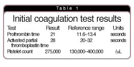
Clinical history. A 75-year-old male who lived alone presented to the hospital by ambulance after being found on the floor in a semiconscious state in his room. A friend stated the patient was not taking medications and had a history of poor nutrition and poor hygiene. Physical exam showed tachycardia, malnourishment, dehydration, disorientation, and guaiac-positive stool. The initial coagulation test results are shown in Table 1.
Laboratory test-based algorithm. Laboratory professionals identified a prolonged prothrombin time (PT) with a normal activated partial thromboplas-tin time (APTT) as the primary coagulation abnormality on the screening tests. The common-and not so common-conditions causing this laboratory abnormality are shown in Table 2.

A laboratory test-based algorithm for systematically evaluating these causes is shown in Fig. 1. This algorithm represents one possible approach for arriving at a correct diagnosis; users can modify it as needed based on the clinical and laboratory information available at the time they first recognize the elevated PT.

The first step is to elicit a history of possible coumarin use in the past seven days. This includes the use of therapeutic coumarin (warfarin) for a medical condition requiring oral anticoagulation, surreptitious coumarin, and coumarin-based rodent poison. If such a history exists, it is reasonable to suspect the elevated PT is due to coumarin effect. If clinically indicated, demonstrating low levels of factor II, VII, IX, and X can further support this diagnosis.
The second step is to evaluate for a liver function abnormality. This can be done by performing tests that measure hepatocellular damage (aspartate aminotransferase, alanine aminotransferase), biliary function (total bilirubin, direct bilirubin, alkaline phosphatase, gamma glutamyl transferase), and synthetic function (albumin, factor V, antithrombin). Acute liver failure from acetaminophen toxicity has been known to present with an isolated prolongation of the PT. This is because of the short half-life of factor VII, which can cause a selective deficiency of factor VII with normal levels of other coagulation factors.
The third step is to consider vitamin K deficiency. Laboratory professionals typically diagnose this by observing the PT correction following administration of vitamin K, plus the presence of clinical risk factors for vitamin K deficiency (poor dietary intake, antibiotic use, biliary tract disease, diffuse gastrointestinal disease, newborn period). If the clinical situation requires the laboratory to distinguish between vitamin K deficiency and liver disease, it is useful to perform a factor V assay and a vitamin K dependent factor assay such as factor VII. A normal factor V and low factor VII supports vitamin K deficiency, while a low factor V and low factor VII supports liver disease.
The fourth step is to consider disseminated intravascular coagulation. If DIC is suspected based on clinical features, then it is useful to measure either the D-dimer or fibrin split product level. If either of these tests is normal, the laboratory can reasonably exclude the diagnosis of DIC as a cause of the elevated PT. If either of these tests is significantly elevated, then DIC is a possible diagnosis, one that the laboratory should further evaluate.
If the cause of the prolonged PT is still elusive at this point in the algorithm, the fifth step is to perform a mixing study of the PT. This test distinguishes between a coagulation factor deficiency and a coagulation factor inhibitor. The test procedure involves mixing equal volumes of patient plasma and normal pooled plasma, then measuring the PT on the mixture. Full correction of the PT into the reference range suggests a deficiency of a coagulation factor within the extrinsic coagulation pathway, such as factor VII. Inherited factor VII deficiency is a very rare condition; laboratory professionals can confirm it by demonstrating a low factor VII level in the patient and a low factor VII level in one or both parents.
Incomplete correction of the PT after mixing with normal plasma suggests two possibilities. The first is a factor VII inhibitor, an exceedingly rare condition. Performing a factor VII assay and a modified Bethesda assay will confirm this. The second possibility is a lupus anticoagulant that selectively prolongs the PT without prolonging the APTT. This is an extremely rare presentation for a lupus anticoagulant.
Diagnosis. The laboratory test results based on this algorithm approach are shown in Table 3. The patient was eventually able to communicate and reported no history of coumarin use. Liver function tests were normal with the exception of a low serum albumin, which supported the clinical impression of malnutrition. Vitamin K deficiency was suspected, and administration of vitamin K rapidly corrected the PT into the reference range, confirming this impression. The negative D-dimer excluded DIC. The 1:1 mixing study of the PT showed correction into the reference range, arguing against a factor VII inhibitor or an unusual lupus anticoagulant. In summary, the prolonged PT was most likely due to vitamin K deficiency arising from malnutrition.

Dr. Cunningham is a member of the CAP Coagulation Resource Committee.