Feature Story |
 |
|
|
Assembly of whole-organ histologic and genetic maps March 2002 
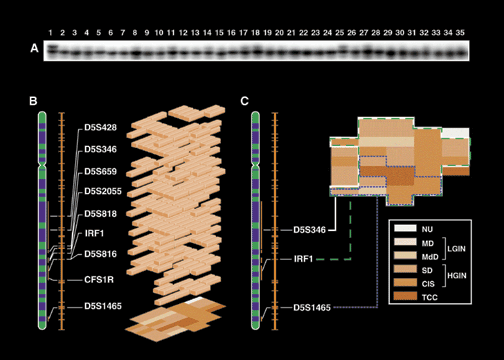
A. An example of marker tested on multiple mucosal samples from the cystectomy specimen (map 5). Marker IRF1 shows loss of heterozygosity (LOH) in samples corresponding to TCC (samples 4, 15, and 16) as well as ones exhibiting changes consistent with LGIN (samples 13, 19, 22, and 27) and HGIN (samples 2, 3, 5-12, 14, 17, 18, 20, 21, 23, 24, 26, 28-35). Sample No. 1 represents allelic patterns of the same marker from peripheral blood of the same patient and serves as control. The presence of LOH in all samples was confirmed by densitometry and is expressed as O.D. ratio below each sample in both panels. O.D.<0.5 is indicative of LOH. B. Example of chromosome 5 allelic losses in a single cystectomy specimen (map 5) with invasive non-papillary urothelial carcinoma assembled by nearest neighbor analysis. The vertical axis represents a chromosome 5 map showing positions of markers and their chromosomal locations. Only altered markers are shown. The shaded blocks represent areas of urinary bladder mucosa with LOH as they relate to progression of neoplasia presented by a histologic map of cystectomy in the background. Note: several markers, including IRF1, show LOH in a form of a plaque involving a large area of urinary bladder mucosa. Code for a histologic map is shown in B. C. Examples of LOH distributions superimposed on a histologic map of cystectomy specimen (map 5). Markers D5S346 and IRF1 show a plaque-like LOH involving almost all of the urinary bladder mucosa. Marker D5S1465 shows LOH involving smaller area of urinary bladder mucosa located within a larger plaque of LOH which involved markers D5S346 and IRF1. Open boxes delineated by lines indicate areas of urinary bladder mucosa with alterations in a given locus. The background shadowed area represents a histologic map of cystectomy specimen depicting distribution of various intraurothelial precursor conditions and TCC. Histologic map code: (NU) normal urothelium; (MD) mild dysplasia; (MdD) moderate dysplasia; (SD) severe dysplasia; (CIS) carcinoma in situ; (TCC) invasive transitional cell carcinoma. Reprinted with permission from Laboratory Investigation (2001;81[7]:1039). |
|||
|
