Feature Story |
 |
|
|
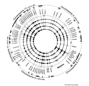
Genetic model of March 2002
Outer circle represents chromosomal vectors aligned clockwise from p to q arms, with positions of altered markers exhibiting loss of heterozygosity (LOH). All the markers are positioned on the vectors according to the human genome database (version March 14, 1996). The innermost concentric circles represent major phases of development and progression of urothelial neoplasia from normal urothelium (NU) through low-grade intraurothelial neoplasia (LGIN) and high-grade intraurothelial neoplasia (HGIN) to transitional cell carcinoma (TCC). Solid circles denote statistically significant LOH of the markers defined by the logarithm of odds score analysis. Open circles identify LOH without statistically significant association to a given stage of neoplasia. The positions of open or solid circles on appropriate concentric circles relate the alterations to a given phase of neoplasia. Only markers with LOH are positioned on the chromosomal vectors. Solid bars on outer brackets represent clusters of markers with significant LOH and denote location of putative tumor suppressor genes involved in urothelial neoplasia. The distances of markers on chromosomal vectors and the solid bars depicting minimal deleted regions were adjusted to fit the circle and are not drawn to scale. More precise localization of these regions can be obtained from individual chromosomal vectors. Reprinted by permission of Wiley-Liss Inc., a subsidiary of John Wiley & Sons Inc. Czerniak B, et al. Genetic modeling of human urinary bladder carcinogenesis. Genes Chromosomes Cancer. 2000;27:392-402. |
|||
|

