 CAP TODAY Pathology/Laboratory Medicine/Laboratory Management

![]()
CAP TODAY and the Association for Molecular Pathology have teamed up to bring molecular case reports to CAP TODAY readers. AMP members write the reports using clinical cases from their own practices that show molecular testing’s important role in diagnosis, prognosis, and treatment. Case report No. 8, which begins here, comes from Danbury Hospital, Western Connecticut Health Network. If you would like to submit a case report, please send email to the AMP at amp@amp.org. For more information about the AMP, visit www.amp.org.
Tsetan Dolkar, MD
Jennifer Zikria, MD
Stuart Bussell, MD
Shannon Morrill-Cornelius, MS, CGC Rina Siddiqui, MD
June 2015—Tumor suppressor genes direct the production of proteins that regulate cell division. Mutations in these genes result in uncontrolled cell growth and may contribute to the development of a cancer. Adenomatous polyposis coli (APC) and breast cancer 2 (BRCA2) are two such genes.
Pathogenic mutations in the APC gene cause familial adenomatous polyposis (FAP), an autosomal dominant inherited disorder, characterized by early-onset colorectal cancer. People with classic FAP develop colonic polyps in their teenage years that eventually undergo malignant transformation. The average age for presentation with colorectal cancer is 39 years. Attenuated FAP (AFAP) is a phenotypically distinct entity, typically presenting with fewer than 100 adenomas. The average age for presentation with colorectal cancer for AFAP is 55 years.1
Heterozygous mutations of the BRCA2 gene can lead to an increased risk of cancers involving the breast in both male and female, ovary, prostate, pancreas, fallopian tube, and skin (melanoma).
 Case presentation: A 44-year-old Caucasian male of Hungarian, Czechoslovakian, and Irish descent with no significant past medical history presented with abdominal pain, distension, nausea, projectile vomiting, and loose stool for a week. He denied fever or chills and had no recent travel or trauma. He also reported unintentional weight loss of 90 pounds in the past two years, with 20 pounds lost in the prior two months. Computed tomography scan of the abdomen and pelvis showed a 3.7-cm “apple core” lesion of the rectosigmoid colon causing large and small bowel obstruction without distant metastasis.
He underwent sigmoid colectomy with end colostomy and ileal colostomy that found a pT4a pN2a M1 colonic adenocarcinoma with 15 additional colon polyps. The tumor was microsatellite stable by immunohistochemistry. Formalin-fixed, paraffin-embedded tumor tissue sent for next-generation sequencing resulted in the diagnosis of a pathogenic somatic KRAS mutation (G12A). Postoperative CEA level was 55 ng/mL (normal 0.0–4.3), which prompted a PET scan that found focal uptake in the right lobe of liver demonstrating SUV uptake 10. The liver lesion was also seen on MRI and underwent biopsy, confirming metastatic colonic adenocarcinoma.
The patient was evaluated by a hepatobiliary surgeon and it was recommended that he proceed with adjuvant rather than neoadjuvant liver resection. He was treated with FOLFOX (FOL—folinic acid [leucovorin], F—fluorouracil [5-FU], and OX—oxaliplatin [Eloxatin]) and bevacizumab (Avastin) for six cycles. At the completion of chemotherapy, MRI and CT scan showed no residual lesions and a normal CEA of 1.1 ng/mL.
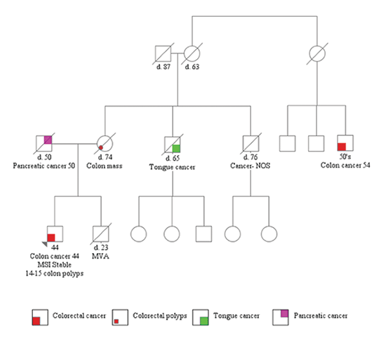
As multiple colonic polyps were seen on colonoscopy, he was referred for genetic counseling. Family history was remarkable for pancreatic cancer in his father at age 50 and colon cancer in his mother and maternal cousin once removed at ages 74 and 54, respectively. The patient is estranged from the paternal side of his family, so that history was unavailable (see pedigree below).
After counseling, he decided to pursue genetic testing. Peripheral blood in EDTA was sent to Ambry Genetics. A panel of 14 genes associated with hereditary colon cancer was tested first by next-generation sequencing and confirmed by Sanger sequencing. The panel included APC, BMPR1A, CDH1, CHECK2, EPCAM, MLH1, MSH2, MSH6, MUTYH, PMS2, PTEN, SMAD4, STK11, and TP53. He was diagnosed with a pathogenic mutation in the APC gene due to a C to T substitution at nucleotide position 1213 in exon 9. The mutation resulted in a premature stop codon instead of the amino acid, arginine (p.R405X) (Figs. 1 and 2).
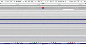
Fig. 1: Results from next-generation sequencing showing a substitution from C to T in the APC gene.
The patient had consented to his DNA being used anonymously for research purposes. The DNA was used to validate BRCA1 and BRCA2 testing for this laboratory. Months after his AFAP diagnosis was made, the laboratory inadvertently unmasked his BRCA1 and BRCA2 results, and his genetic counselor was contacted and asked if the patient would be interested in having those results. The patient consented, blood was drawn for clinical confirmation of the research result, and the patient was informed that he has the deleterious c.8297delC alteration in the BRCA2 gene (Figs. 3 and 4).
He ultimately underwent colostomy reversal. Unfortunately, seven months after colostomy reversal his CEA levels began increasing, rising to 38 ng/mL, and CT scans found a local recurrence at the anastomosis site and three liver lesions. He then underwent complete proctectomy because of the AFAP mutation and excision of the liver implants. He is currently receiving adjuvant chemotherapy with 5-FU and Avastin. Because of the AFAP mutation he is being followed by routine surveillance with upper endoscopy, and thyroid examination. For his BRCA2 mutation, he requires surveillance with annual PSA screening, mammography, and ophthalmologic evaluation.
Discussion: Colorectal carcinoma is one of the leading causes of cancer death in men and women in the United States. While significant progress has been made in the treatment of metastatic colorectal carcinoma, one of the major challenges is resistance to chemotherapy treatment. KRAS is an oncogene that functions downstream to membrane-bound receptors such as EGFR. Mutations in KRAS result in a constitutively activated protein leading to unregulated proliferation, impaired differentiation, and resistance to EGFR inhibitors.2 The G12A point mutation activates KRAS and is associated with resistance to EGFR tyrosine kinase inhibitors (erlotinib, gefitinib) and monoclonal antibodies (cetuximab, panitumumab).3–5
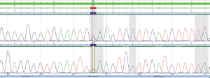
Fig. 2: Chromatogram of Sanger sequencing confirming the findings by next-generation sequencing.
Friedl, et al., noted that alternatively spliced sequence of exon 9 and mutations in 5′ to codon 168 and 3′ to codon 1580 were associated with AFAP phenotype.6 A review of the literature revealed one case report in German that described the same mutation our patient had in an asymptomatic 59-year-old female with multiple tubular and tubulovillous adenomas on routine colonoscopy.7 Additionally, Soravia, et al., characterized mutations located in exon 9 in two kindred with AFAP: one with deletion of nucleotide ‘A’ causing frameshift and early stop codon at nucleotide 1014 and the second with C to T substitution resulting in a nonsense mutation at nucleotide 994.8 Therefore, the mutation found in our patient can be assumed to be associated with the AFAP.
The deleterious c.8297delC alteration in the BRCA2 gene is a pathogenic mutation located in exon 17 of the BRCA2 gene.9 It causes a translational frameshift with a predicted alternate stop codon. The BRCA2 protein is part of a complex that repairs homologous double-stranded breaks through the interaction with RAD51. Thus,