 CAP TODAY Pathology/Laboratory Medicine/Laboratory Management

Bridget Kuehn
October 2014—Like many cytopathology trainees, Charanjeet Singh, MD, who recently completed a cytopathology fellowship at MD Anderson Cancer Center in Houston, found it challenging at times to find classic examples of entities to learn from and to study for exams. Most texts he consulted contained just one or two images of a particular diagnosis. And the material in training programs from all specialties varies. Even though there is a large volume of cytology cases at MD Anderson, for example, it wasn’t enough to learn gynecologic cytology, which is why he pursued an elective rotation at Houston Methodist Hospital.
“I was struggling to find good examples of cases to learn from,” Dr. Singh says. “I wondered how many more residents and fellows were in the same boat.”
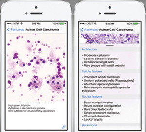
Users can view multiple photographs for each diagnosis and detailed descriptions of each image.
During his fellowship, Dr. Singh shared his frustrations with his mentor, Sinchita Roy-Chowdhuri, MD, PhD, an assistant professor of pathology at MD Anderson. As a trainee, she too had been frustrated by the lack of convenient options for studying cytology. Most slides weren’t available when she needed to study, she says, and carrying multiple bulky cytology texts or atlases wasn’t feasible.
They both agreed that a mobile application would be an ideal solution to this problem, but none existed. So over the course of several months, the pair teamed up to create one.
“Most of the time when you study, you don’t have many books or a microscope,” Dr. Roy-Chowdhuri says. “But everyone has a smartphone.”
The fruit of their labor is CytoAtlas, a free mobile application that debuted in the iTunes store in August. Dr. Singh will present the app at the American Society of Cytopathology’s annual meeting Nov. 14–17 in Dallas.
The app, which is available now only on an iOS platform, has more than 750 reference images for more than 100 different diagnoses. Students can review multiple photographs for each diagnosis along with detailed descriptions of each image, including information about the architectural, cytological, and nuclear features that distinguish each disease entity. Information is also available about the organs and systems affected by the disease entities. The cloud-based app is searchable by diagnosis.
To build the app, Drs. Singh and Roy-Chowdhuri began selecting disease entities in March. For each entity, they took 30 to 40 photographs using a high-quality camera attached to a microscope. From this huge catalog of photos, they selected the best images of each disease entity and included them in the app. They wrote descriptions for each entity and collected relevant citations from the medical literature. It took five months to amass the content.
But neither had the technical skills to write the computer code necessary to build the app. So they hired ProQMS, a Web development and information management company, to build the app itself (www.proqms.com). ProQMS built the app over a three-month period, as the content for the app was being collected.
The iPhone app has been updated for iOS 8, the latest operating system, and a universal app for both iPhone and iPad is in development and will be available soon. Dr. Roy-Chowdhuri says they chose to start on the iOS platform because 80 percent of U.S. medical trainees have a smartphone, and the iPhone is the brand of choice for more than half of trainees (Franko OI, et al. J Med Syst. 2012;36:3135–3139).
The app quickly found an audience. Within weeks of its launch, more than 100 copies had been downloaded, Dr. Roy-Chowdhuri says. At that time, she and Dr. Singh had done little to promote the app, which suggests, she says, that many students and pathologists were actively searching for a cytology app.
For now, cytotechnology students, pathology residents, and fellows studying cytopathology are the primary audience for CytoAtlas. Dr. Singh notes that in 2012, about 2,200 residents studied cytology for at least three months at U.S. training programs, and more than 140 individuals entered fellowships.