 CAP TODAY Pathology/Laboratory Medicine/Laboratory Management

Karen Titus
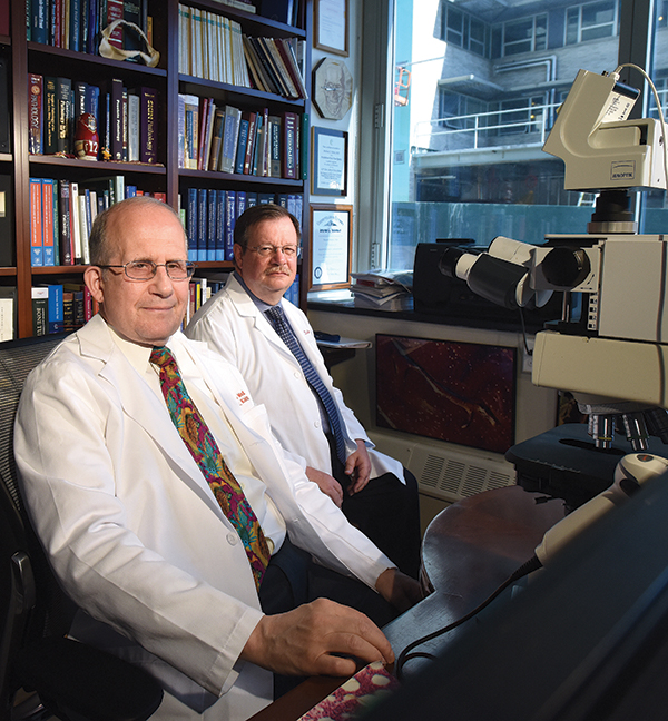
Dr. Michael Klein (left) and Dr. Edward DiCarlo
made a compelling case in 2014 for comparing clinical and histologic diagnoses in patients
undergoing total joint arthroplasties.
July 2017—One of the more unnerving scenes in contemporary theater comes courtesy of Martin McDonagh’s “A Skull in Connemara,” which opens with two men in an Irish graveyard, hired by the local priest to make room in the overcrowded burial ground. Their method? Exhume the corpses and smash the bones to bits.
The action is macabre and outrageous. Practically speaking, it’s also efficient (and in McDonagh’s hands, distressingly funny). What else can one do with bones that don’t seem to matter, that are old and now in the way?
A similar question taunts pathologists and surgeons. When performing total joint arthroplasties, what should surgeons do with the hip and knee specimens? Submit them for pathologic examination? If so, is a gross examination sufficient? When might a microscopic examination be in order? Is it ever okay to channel McDonagh’s Irishmen and toss the bones aside as (biohazardous) rubbish?
A look at the orthopedic literature suggests the latter approach might actually have fiscal soundness. The message is clear from titles such as: “Histologic examinations of arthroplasty specimens are not cost-effective” (Lin MM, et al. Clin Orthop Relat Res. 2012;470[5]:1452–1460). The point of these studies, generally, is that routine pathologic exams boost costs but rarely alter patient management. The phrases “limited cost-effectiveness” and “low prevalence of findings” pop up with the regularity of a president on a golf course.
The reception these studies receive from pathologists, however, can be chilly.
Take, for example, a letter spearheaded by James Richard, DO, who responded to a study, which took place at the institutions where he practices, that suggested histologic examinations of shoulder arthroscopies did not influence patient care or provide new diagnoses. In the letter, published in the Journal of Bone & Joint Surgery in November 2008, Dr. Richard, who is director of laboratories, Sparrow Health System, Lansing, Mich., argued the value of doing such exams. In one 12-month period alone, he and his colleagues wrote, they uncovered nine cases of malignancy or probable malignancy from the examination of all orthopedic specimens (not just shoulder arthroscopies).
Dr. Richard’s views remain undimmed nearly a decade later, in 2017. Sparrow does not routinely do microscopic exams. “We’re not even receiving the specimens,” he says.
Dr. Richard says the decision was made before his arrival. Bringing it up for discussion “would be opening Pandora’s box,” he says. At another institution where histologic exams were routine, he had to defend the practice on more than one occasion. He succeeded, but since his departure, he notes, the institution has stopped sending total joint specimens to pathology. He sounds a bit weary as he weighs his efforts over the years. “I’ve been tilting at that windmill for quite some time.”
Keep tilting, says Michael Klein, MD. With a 16,587-specimen study to back up his points (DiCarlo EF, Klein MJ. Am J Clin Pathol. 2014;141[1]:111–118), Dr. Klein makes a compelling case for comparing clinical and histologic diagnoses. As he points out, most previous studies top out at 1,200 or 1,500 cases. “And those series are usually collected from a couple of hospitals and combined,” says Dr. Klein, pathologist-in-chief emeritus, Department of Pathology and Laboratory Medicine, Hospital for Special Surgery, New York.
Dr. Klein, who is also a professor of pathology and laboratory medicine, Weill Cornell Medical College, and consultant in pathology, Memorial Sloan Kettering Cancer Center, sees more than his share of total arthroplasty specimens. At Special Surgery, there may be 40 to 60 total joint replacements a day. The topic is inherently relevant to his work, but he says his interest has also been provoked by the extensive orthopedic literature that suggests certain pathology—and even certain radiology and indeed surgical—procedures are not considered to be cost-effective and thus should not be done.
“It all sounds very altruistic,” he says. “It’s not exactly altruistic. I believe a little bit of it is self-serving and self-preserving, which I understand.” Moreover, he says, the orthopedic literature often neglects to point out reasons other than cost-effectiveness for doing these exams, including quality assurance, risk management, and, of course, patient care.
Dr. Klein also bristles at study authors who draw their conclusions based only on whether histologic exams identify a clinically unsuspected tumor. This is a fallacy, he says, since tumors are relatively rare anyway, and a careful gross examination is needed before a pathologist decides to take sections. A microscopic examination can identify tumor type, but it’s not needed to find the tumor to begin with.
Spurred by what he saw as the lack of useful studies, Dr. Klein and Edward DiCarlo, MD, undertook their own study, assessing total joint replacement specimens (7,968 hips, 8,619 knees) over a 10-year period that had been grossly and microscopically examined by Dr. DiCarlo (chief of surgical pathology at HSS) and verified by Dr. Klein.
Notably absent from their study was an attempt to provide cost analysis. The pathologists simply wanted to compare the postoperative surgical diagnosis with the pathologic diagnosis for the seven most common diagnoses: degenerative joint disease, traumatic injury/fracture, avascular necrosis, subchondral insufficiency fracture, rapidly progressive arthritis, inflammatory arthritis, and septic arthritis. Dr. Klein says they expected their study to mirror earlier orthopedic studies, which found no significant differences between clinical and histologic diagnoses.
The numbers told a much different story. The discrepancy rate was 18.8 percent for hips and 9.4 percent for knees. And 5.4 percent of hip joints and 1.4 percent of knee joints showed discordant histologic findings that were clinically unsuspected and should have affected patient management and outcomes.
Surgeons who say examinations aren’t needed because their clinical assessments are correct are, well, wrong, says Dr. Klein. There are, he says, important diagnostic findings apart from tumors, particularly subarticular insufficiency fractures. This diagnostic category is often mistaken for degenerative joint disease, but should be treated differently. “In fact, because its clinical history is different from degenerative joint disease, it should probably be diagnosed preoperatively by surgeons, and it’s not,” he says. In some cases, a total joint replacement may not be merited as a primary treatment. In fact, this condition is often associated with underlying comorbidities, such as morbid obesity, metabolic bone diseases, and lifestyle choices that may be altered with appropriate medical advice and treatment to prevent future fractures in other joints.
Degenerative joint disease was the most common diagnosis among surgeons and pathologists, but familiarity didn’t necessarily breed accuracy. It was also the most overdiagnosed condition—it was diagnosed approximately 20 percent more frequently than could be verified by the pathologists. And, the authors say, despite its prevalence, it was not always recognized clinically when it was present.

Dr. Bachner
Paul Bachner, MD, sings the praises of the Hospital for Special Surgery, noting its status as a highly acclaimed orthopedic hospital. “You have to assume their orthopedists are extremely experienced.” Nonetheless, the study uncovered those 18.8 and 9.4 percent error rates. “And this is a group that’s probably as expert as you can find in the country,” says Dr. Bachner, a professor of pathology and past chair of the Department of Pathology and Laboratory Medicine, University of Kentucky, Lexington.
(The flip side, he says, is that Drs. Klein and DiCarlo are two of the best orthopedic pathologists in the country. “Their ability to find things may be greater than a pathologist who does not specialize in that area.” As they themselves noted in their study, the two had an aggregate experience in bone disease analysis of more than 65 years.)
In his own experience, Dr. Bachner, who in June retired as director of laboratories at UK, says the most common finding is avascular necrosis in cases where there’s a radiologic and clinical diagnosis of osteoarthritis. “And every once in a while you’ll find a cancer—it’s very rare.”
Dr. Klein has plenty to say about histologic exams of total joint arthroplasties. But he’s succinct on one point in particular: Don’t abandon histologic exams because of so-called cost-effectiveness. “Cost-effectiveness isn’t the reason we do pathology.”
Another reason to do histologic examinations, Dr. Klein says, is to learn what the actual disease rate is in a series of surgeries on a site for a particular surgeon, just as one would for any organ system. Before the advent of CT scanning, the acceptable excision rate of normal appendices for clinically suspected acute appendicitis was 15 percent. Significantly more than this and a surgeon was too aggressive; significantly less would mean the surgeon was too conservative. Is there an acceptable rate for removing histologically normal joints? “While this ideally should not happen, there is probably some very small finite percentage that is permissible,” Dr. Klein says. He has no idea what that number is, but determining the rate is important. “If you throw that information out, then you’re neglecting an essential part of public health.”
Dr. Richard links the matter to the broader picture as well. He’s familiar with the arguments that a bone might have broken based on circumstances—say, an auto accident. “But was there anything causing a weakening for it to be broken at that specific site, that specific bone? Was there an unknown defect that caused it?” That’s just as true for hip joints, he says. “How many times do we hear in the literature about patients falling and fracturing a hip?” Pathology might reveal an occult malignancy; it might also reveal metastatic spread. If the pathologist finds crystals, it may be indicative of a systemic disease. “There’ve been times when we’ve picked up acute inflammation” indicating possible sepsis. “And there are times when you find what might be considered subtle changes, but they aren’t—they’re diagnostic.” The obvious, in short, isn’t always obvious. “It goes back to the diagnosis and documentation that are part of our role.”
Dr. Richard urges pathologists to take what he calls the higher road: concern about patient care and patient safety. “That’s where we’ve got to hang our hat.” At the minimum, pathologists should look at all fracture cases, he says, or any cases the surgeon deems unusual.
“We’re guardians of the galaxy here, if you will. We’re trying to prevent potential threats, hoping they never come to fruition.” — James Richard, DO
“I would encourage, for documentation purposes, all specimens be submitted,” he continues, “but a majority of them would be for gross-only evaluation.” Microscopic evaluations would occur only if the pathologist thought the case was unusual enough to merit a closer look, or if the surgeon requested it, based on the patient’s history and clinical information. “Most of us in pathology recognize standard degenerative changes within that joint surface,” Dr. Richard says. “We see it, we document it, we put out a simple report saying, ‘Yes, it is consistent with that.’ And that would be the end of it.”
Such efforts are, he says, the equivalent of following the rules of the road. A driver at an empty intersection stops at the stop sign, he says, “even if it’s in the middle of the Utah desert and you can’t see anyone for miles in any direction. This is what you do.”
So why might some physicians be tempted to blow through that stop sign? Perhaps the answer has something to do with the dollar sign.
Dr. Klein suspects that if payment is perceived to be at the heart of the matter, pathologists will find it hard to bring up the issue. “Because why all of a sudden are we so interested?” he asks. Surgeons might assume it’s because pathologists want to collect a professional component. “Why else would we care?”
In the case of bundled payments, money for the pathology work comes out of—to put it crudely—the surgeon’s pocket. Although the pathology payment might be small, some surgeons refuse to part with it, says a pathologist whose pleas to surgeons and administrators to allow histologic exams of total joint fell on deaf ears. “No one wanted to give up any portion—in this case, $100—of the bundled payments to the lab. So now the surgeons just throw the hips away.”
Pathologists have financial incentives, too, of course. In smaller practices especially, Dr. Richard says, “Every little case is important financially.”
If pathologists are questioned about their financial incentives, Dr. Richard has a simple suggestion: Offer to cut your fee by 10 percent for a gross-only exam if the surgeon will cut their fee by the same percent. That should put a quick end to the discussion, he jokes, and bring the focus back where it belongs.