Karen Titus
January 2017—Only a sadist would want to see gastroesophageal adenocarcinoma become as common as breast cancer. GEA wreaks enough destruction already as the fifth (stomach) and eighth (esophageal) most common cancers worldwide.
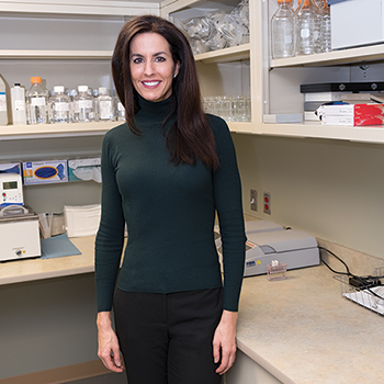
Dr. Angela Bartley, lead author of the HER2 GEA guideline, says she and colleagues concluded from a review of the literature that using biopsy or resection specimens for testing is acceptable. “And if these two are not available, an FNA specimen can be used from the primary or metastatic tumor.”
“It will never be as common as breast cancer—well, I hope it will never be as common as breast cancer is now,” says Mary Kay Washington, MD, PhD, co-chair and coauthor of a new CAP/ASCP/ASCO guideline on HER2 testing in GEA.
Nevertheless, it’s nearly impossible to consider the role of HER2 in GEA without thinking about breast cancer—not with envy, surely, but with an odd covetousness, knowing that accompanying breast cancer’s many more cases are many more studies, and, as a result, much deeper knowledge of how and when to test for the biomarker. With GEA, many mysteries still linger.
But now when pathologists and their clinical colleagues assess HER2 in patients with GEA, they will be better prepared for what they see and how to respond. The new guideline (Bartley AN, et al. Arch Pathol Lab Med. 2016;140[12]:1345–1363) should, say its three co-chairs, be useful to general pathologists as well as those with GI expertise, and to medical oncologists in community and academic settings.
Whatever information had been available to physicians was fragmented, scattered throughout the literature. “This guideline brings everything together,” says Jaffer Ajani, MD, guideline co-chair (representing the American Society of Clinical Oncology) and coauthor.
Many physicians “don’t know a whole lot about this subject as individuals,” continues Dr. Ajani, professor of medicine, and member, Department of Gastrointestinal Medical Oncology, University of Texas MD Anderson Cancer Center, Houston. “Their experiences are based on their work in breast cancer and the literature on breast cancer.”
Until recently, the most widely understood piece of knowledge about HER2 testing in GEA could fill a shot glass: It is the only biomarker established for selecting trastuzumab (Herceptin) to treat advanced GEA.
But then what? For clinicians in particular, “We’re always wondering, should we get a biopsy from a metastatic site, or get a lymph node, or should we go to the primary? This has not been clear,” Dr. Ajani says.
Angela Bartley, MD, another co-chair (representing the CAP) and lead author, adds other questions. Should labs perform immunohistochemistry and in situ hybridization simultaneously? Should pathologists use specific antibodies or probes? When validating a test, how many specimens should be used? Can they be breast specimens, or must they be GEA specimens?
And on the most basic level, “Who should be tested?” asks Dr. Washington (American Society for Clinical Pathology co-chair), professor of pathology, Vanderbilt University Medical Center, Nashville, Tenn.
For some, the answer to that last question is nothing short of critical. It’s no secret that GEA has a poor prognosis, with disease often diagnosed at an advanced stage. As the guideline’s authors note, at this point in a patient’s status—unresectable local-regional, recurrent, or metastatic disease—therapies are limited.
The well-known Trastuzumab for Gastric Cancer Trial (ToGA) stirred a vigorous dash of hope into matters, showing that Herceptin prolonged overall survival compared with chemotherapy-only regimens in those with HER2-positive advanced GEA. Those with IHC scores of 3+ experienced more benefit than those with IHC of 2+ (and concurrent HER2 ISH amplification).
A trio of recommendations from the new guideline reinforce the clinical implications of the ToGA trial. Treating physicians should request HER2 testing on tumor tissue for patients with advanced GEA who are good candidates for combination chemotherapy plus trastuzumab. “It’s kind of a simple recommendation,” says Dr. Bartley—but an important one. Physicians can also banish concerns about trastuzumab’s side effects, the guideline notes. In the ToGA trial, the adverse cardiac event rate was six percent and did not differ between the treatment groups. Patients who received Herceptin had slightly higher rates of other events (including diarrhea, anemia, thrombocytopenia, fatigue) though frequency of side effects was the same.
The question most pressing to Dr. Ajani and fellow clinicians was, what sort of specimen should be tested? “We went through and exhaustively looked at the literature, and determined that using biopsy or resection specimens is acceptable,” says Dr. Bartley, who is the division chair of pathology and laboratory medicine, section head of molecular diagnostics, and gastrointestinal pathologist, St. Joseph Mercy Hospital, Ann Arbor, Mich. “And if those two are not available, an FNA specimen can be used from the primary or metastatic tumor.” A (limited) number of studies suggest a fair amount of concordance between primary and metastatic tumors, she says.
It’s worth noting that resections aren’t always available in GEA cases, though hopes had been high for using this type of material. The thinking was that since gastroesophageal tumors have great heterogeneity compared with breast, says Dr. Bartley, resection specimens would be better in GEA testing, “because we’d have more neoplastic tissue to evaluate.” As it turns out, “The biopsy specimens correlate pretty well with resection specimens.” Since biopsies are more readily available, this is good news.
That’s not to say doing a biopsy guarantees adequate material. “It’s absolutely imperative that you talk to your gastroenterologists or surgeons to get an adequate specimen,” Dr. Bartley says. “The chance of that occurring is, obviously, higher if you communicate with them than if you don’t.” The new guideline doesn’t provide a recommendation but notes that one study recommends testing at least five biopsy fragments of tumor, and the National Comprehensive Cancer Network and other guidelines recommend six to eight. Ideally, she says, clinicians are reading the same guidelines as pathologists and are familiar with these optimal numbers. “But it doesn’t always happen,” she concedes. “Not every institution has a GI advisory board.” (Hers does.)
As the guideline moves to pathology-specific recommendations, including the testing algorithm itself, a theme emerges. Were Chicago Cubs manager Joe Maddon to turn it into a slogan for one of his infamous T-shirts, it might read, “Try not to miss.”
The best way to identify patients who are likely to benefit from trastuzumab is to perform IHC testing first, followed by ISH when IHC results are 2+, or equivocal. “If the IHC is 0, 1+, or 3+, you’re done,” Dr. Washington says.
But recalling the early, unsettled days of qualifying breast cancer patients for Herceptin, when there was pressure to test by all possible means, this straightforward approach for GEA might create flickers of doubt for some. Why not test every specimen using IHC and ISH, just to be sure?
Today, there’s no shortage of data on qualifying breast cancer patients, but the GEA data are less plenteous. The authors had two randomized controlled trials on which to base their algorithm recommendation. That could sow uncertainty: “Some people are thinking, ‘Oh, you’re going to miss people who would be candidates for trastuzumab if you don’t do concurrent IHC and ISH,’” Dr. Bartley says. Especially since there’s evidence—though there’s contradictory data as well—to suggest some negative IHC samples might be positive on ISH.
Based on the ToGA trial, however, it appears that those who were negative by IHC, i.e. 0 or 1+, did not respond to trastuzumab even if they were ISH amplified. “So that’s why for now we’re recommending that people do IHC first, and then do ISH only in examples of 2+/equivocal cases of IHC,” Dr. Bartley says.
It’s hardly a secret that laws often are created in response to what’s happening (as well as to what some fear might happen). And while a practice guideline is hardly law, it’s fair to assume that at least a small percentage of laboratories are performing IHC and ISH on all specimens, rather than waiting to do the latter only on 2+/equivocal specimens.
At Dr. Bartley’s institution, pathologists follow the new guideline “to the letter,” she says. This includes doing IHC first, on all specimens, and following up with ISH on 2+ specimens.
The clinicians cede GEA HER2 testing decisions to the pathologists. That’s put her and her colleagues in the unusual position of not having to communicate closely with clinicians. “I can honestly say I have never had a call from a clinician questioning a result or asking if we should do something else.” Lest some poor business consultant’s head implode at this revelation, she adds, “It’s probably the only testing situation I can think of that clinicians don’t call us and say, ‘Hey, what’s going on with this?’ ”
Some physicians might have hoped the guideline would “do more,” as Dr. Washington puts it. “We know,” she says, “that there were advocates for doing FISH on everything, along with IHC. But I think overall, when you look at the data, most people were happy to accept the final guideline.”
Reflex testing for every patient with esophageal, gastroesophageal junction, or gastric adenocarcinoma is not recommended. “Many of those patients are not recommended for HER2-targeted therapy,” Dr. Washington says. The guideline gives physicians leeway in deciding how and when to test.

Dr. Washington
Dr. Washington’s institution also does reflex testing on 2+ cases, which was instituted at the request of the medical oncologists several years ago. “We will test biopsies or resections or whatever we have the first time we see one of these specimens.”
For a long time they’d done both ISH and IHC on all specimens. The concern was less about missing patients and more about understanding the intricacies of IHC in GEA, with its different scoring system, she says. “We wanted to get a feel for what an amplified case might look like by IHC. There are great published examples of 3+, 2+, but then when you actually start to evaluate one, sometimes it’s not as easy. So that helped us learn about these tumor types.” Some of her colleagues still do both. “I don’t fault them at all,” she says, though she herself has taken to using the guideline’s algorithm. “If I see a 3+ case, I don’t see any need to do in situ hybridization.”
Vanderbilt has a separate GI pathology service, with five GI pathologists and two general pathologists who rotate in. “We have policies and procedures for our little practice that we ask the general pathologists to follow,” Dr. Washington says. “We have tight control over this—everybody knows the expectations.”
Interestingly, the breast pathologists at Vanderbilt also perform ISH for GEA specimens. When the testing first came on board, Dr. Washington and her GI colleagues sat down with the breast pathologists to review cases. “Everybody looked at cases together. We put together study sets and just looked at them.” That was matched by plenty of presentations in multidisciplinary conferences, which included pointed discussions with medical oncologists about how they were using trastuzumab in GI cases.
“But none of us GI people felt we would do it often enough to keep our skills up,” she says. They continue to work closely with the breast pathologists doing the ISH—the GI pathologists give the IHC and H&E slides to the breast pathologists and often go over the areas of concern. “We have to make sure that they’re looking at the most amplified areas and that it’s invasive carcinoma. We don’t want people scoring in situ disease.”
The reflex route is appealing to Dr. Ajani, who says interacting with other members of the guideline committee opened his eyes to new possibilities.
“I think the ordering should be standardized,” he says. At MD Anderson, the electronic medical record doesn’t always alert him to a 2+ IHC result in a timely way, and he says he wastes additional time having to order FISH on such specimens. “If pathologists are about to report a 2+, they should themselves order the FISH,” he says. “That will save several days, because they know I’m going to order it anyway.”
He’s not alone in his thinking—this topic came up in the guideline discussions. But that’s as far as matters got. “There was no agreement,” he says. For that to change, he suspects, pathologists, with their varying practice patterns and cultures across institutions, would need to be the ones to insist on a single algorithm. “Clinicians would welcome them to proceed with certain automated testing,” he contends, “because we know we’re going to need it.”
Other parts of the guideline are less likely to spur lengthy barroom discussions among pathologists.
The guideline is agnostic on the question of using specific antibodies or probes. Rather, pathologists need to document what they’re using and validate the assays for IHC and ISH on GEA specimens. “There simply weren’t enough data to help choose one antibody over another. So that’s very laboratory dependent,” Dr. Washington says.
It does get more specific about other aspects of validation. “For FDA-approved tests, we’re recommending you use 20 positive and 20 negative specimens,” says Dr. Bartley. For laboratory-developed tests, the recommendation is 40 of each. Perhaps not surprisingly, “This is pretty much what the breast guidelines recommend.” Also not surprisingly, there are few studies looking at validation in GEA testing. “So we had to kind of borrow from the breast guideline,” she says.
In the early days of absorbing the ToGA data, there was hope that pathologists’ experience with breast cancer and HER2 testing would translate almost directly to GEA, including validation. That dream has died.
“We realized,” Dr. Bartley says, “that for most institutions, the proportion of breast samples—whether it’s resection or biopsy—is greater than what you would have for GEA.” Some laboratories have found it difficult to obtain 40 or even 20 positive and negative specimens, especially since not many GEA cases are actually positive for HER2. “So we’re aware that some places have done their HER2 validation on breast only,” and possibly trying to throw in a few GEA specimens for good measure.
“However, we’re recommending, when possible, to use GEA specimens in your GEA validation,” she says firmly. “We’re advocating using as many as possible, because of the differences in heterogeneity.”
The supply issue points to another concern. Dr. Bartley’s practice includes a small cadre of GI pathologists as well as other specialists; there are only a few generalists. “So only a few of us read HER2 for GEA. As we stress in the guideline, you have to keep up your skills for this, and it takes practice.” Though St. Joseph is a community practice, “We see a fair amount of volume of GEA.” In fact, they had enough tissue to do a full, 40-sample validation for GEA.
But not every pathologist is in such a position. For those who see fewer cases and may not be able to attain or maintain expertise in HER2 interpretation, it might be worth considering sending out the test to ensure patient safety, Dr. Bartley says.
The guideline recommends the Ruschoff/Hofmann method for scoring IHC and ISH results, providing examples, color photos, and—naturally—a discussion of breast cancer. Similar to breast cancer, HER2 scoring for GEA relies on membranous staining but not cytoplasmic staining. Unlike in breast cancer, however, GEA does not require complete membranous staining; incomplete or basal lateral staining is sufficient. The authors explain that often the luminal surface of tumor cells fails to stain in HER2-amplified GEA, and that only luminal surface staining in the absence of lateral and basal staining should be considered negative.
“Pathologists really need to know that the immunohistochemistry is different for these GI cancers than it is for breast cancer,” says Dr. Washington. “So we cannot apply the breast criteria.”
Dr. Washington hopes the guideline torpedoes any lingering misperceptions about possible similarities. “Early on there may have been a problem there,” she acknowledges. But since the ToGA trial has been out for several years, “I’m hoping that pathologists have gotten the message.” The problem with using the breast criteria is that they exclude too many people—maybe 25 percent or so—who would otherwise be eligible using the GI criteria.
Pathologists should select the tissue block with the areas of lowest grade tumor morphology. The rates of HER2 positivity in GEA is greater in the lower grade tumors than in higher grade tumors, Dr. Bartley explains, as well as at the gastroesophageal junction versus the stomach.
And if ISH is required, pathologists need to mark the area of strongest intensity for IHC HER2 expression. If, for example, the pathologist can choose from among a 2+, 1+, and a negative area, the 2+ area needs to be circled for testing on an equivocal case, Dr. Washington says.
Similar simplicity shapes the guidance on templates. Every laboratory has and can use its own template for reporting HER2 results, Dr. Bartley stresses. And while they don’t need to follow the CAP template word for word, it would be wise for them to examine it to make sure all the elements are there. Labs also need to include laboratory quality improvement in their validation.
The past few years have seen a considerable uptick in the information about breast and HER2, some of which, as noted, has filtered down to the GEA guideline. But Dr. Bartley wants more. “I’m hoping that there are a lot more studies in GEA in the future. Because it really is needed,” she says.
Ideally, this would include a large, randomized controlled trial looking at patients who are negative by IHC and positive by ISH. In the ToGA trial, HER2-positive results by FISH were observed in 11 percent of cases with an IHC score of 0 and in 12 percent of cases with an IHC score of 1+. But absent larger trials, the evidence gap resembles those in the fossil record. Greater numbers would lay to rest questions about the response of patients who are ISH amplified.

Dr. Ajani
Dr. Ajani expresses disappointment that the guideline leaves unresolved the number of specimens that need to be tested from a single patient. “This is entirely unclear,” he says.
Traditionally, that number has stood at one. “When I request HER2 testing, the pathologist will just take one section, one slide, probably from a block, and make sure there’s tumor there,” Dr. Ajani says. “They may also find a more differentiated area, stain it, and that’s the end of it.”
But, he continues, when he and his clinical colleagues inadvertently request repeat HER2 testing (for example, on outside cases where HER2 testing initially appears not to have been done), “the two results can be totally different—3+ on one result and 0 on the other,” thanks to the heterogeneity that characterizes GEA specimens. “So nobody has come up with an answer on how many samples we should study.”
He’d like to see a robust study done—approximately 100 patients, with four to six specimens per patient, from one or two blocks—to assess discordance. Some limited work in this area has already been done. “But we have to finish the research, and if the results really change our understanding, that has to be implemented,” he says.
Based on his own experience, he remains concerned about inter-pathologist discrepancy on IHC reads. “I’ve noticed there is a lot of disagreement among pathologists on 1+ and 2+. That is worrisome to me.”
Also sorely needed: further studies in both GEA and breast on the meaning of polysomy with regard to response to trastuzumab. For now, the GEA guideline once again takes a page from the breast guideline, where there is slightly more (but still not much) information.
When the guideline group was putting the document together, Dr. Bartley recalls, “We had a lot of questions on polysomy while we were still learning over the last two years and putting the data together.” She even received personal emails from those in the group, asking what she’d found on the topic. “There’s not a lot out there,” she laments.
There’s a paucity of data on turnaround time and other technical issues related to HER2 testing for GEA. A 10-day TAT is fairly standard, but it’s hardly popular with clinicians, Dr. Ajani says. Calls for a faster TAT came up in the guideline discussions as well, he says, with an initial suggestion of three business days. “There was a lot of objection to that.” But the 10 days that made it into the guideline is too long, he says, and pathologists who can reduce that will be helping their clinical colleagues tremendously.
For tissue fixation, the guideline makes recommendations that echo what’s done in breast and other tissue. But there’s very little in the literature about ischemic time to fixation for GEA. It would be nice to have more data on fixation and other preanalytical variables, says Dr. Washington, but she’s not holding her breath. “Most places in this country don’t see that many gastric resections, so there’s not much material to work with.” Japan and Korea, where the procedure is more common, might be the source of these studies. Similarly, she’d love to see more studies looking at those thorny subgroups—IHC-/ISH+ cases, for example—but questions whether enough such cases will ever be compiled for a randomized prospective clinical trial.
Ditto for polysomy. “Our problems are case numbers,” Dr. Washington says. The United States sees a fair number of esophageal adenocarcinomas, but fewer gastric cancers. The relevant cases drop drastically when HER2 positivity is considered, and the numbers get even smaller with the rare subgroups.
Given the lack of published data on GEA, this may not be a guideline for the ages. But rest assured, says Dr. Washington, the guideline is also based on expert consensus, not to mention institutional expectations, good laboratory practice, and good medical care.
Adds Dr. Ajani, “This is going to be a helpful document.”
[hr]
Karen Titus is CAP TODAY contributing editor and co-managing editor.