Julietta Fiscella, MD
January 2014—Meaningful use standards are fostering increasing patient access to medical records,1 including pathology reports. Yet pathology reports can be challenging even for clinicians,2 much less for patients, to understand.3 Nonetheless, it is typically left to the treating non-pathologist clinician to explain the findings to the patient, even when the clinician lacks detailed knowledge of pathologic features.
Patients are becoming better informed consumers of health care. Aided by ready access to Web-based health information and pathology images, many patients use the Web to research their disease and view pathology images.4 While Web sites, such as the CAP’s MyBiopsy.org, can be helpful,5 there is no substitute for expert, personalized explanations of pathology for patients.6
One way to address the growing need patients have for expert explanation of the pathology diagnosis is to foster direct communication between pathologists and patients, particularly when the pathology is challenging.3,7,8 Yet despite calls dating back more than 30 years for direct communication between surgical pathologists and patients,9 little is known of such communications, and I am not aware of published accounts. Here, I describe my experience with a pilot project in which a surgical pathologist meets with selected patients at the bedside and reviews and explains the pathologic findings with the patient and family.
We adopted a multidisciplinary team approach to reviewing pathology reports. The team consists of the surgical pathologist, the attending hospitalist physician, medical residents, and medical students at a university medical center.
The attending hospitalist physician offers patients the opportunity to discuss their pathology reports and to view and discuss their pathology images with the team. He selects those who have a diagnosis that is challenging to understand and are likely to have an interest in their pathology images. The project began in 2011 and was designed as a clinical program, not a research project, and is exempt from institutional review.
As the surgical pathologist who meets with these patients, I prepare for the consultations by creating digital images at varying magnification. I also prepare corresponding, normal histological images to contrast with the patients’ abnormal findings. My consultation with patients takes place within a hospital conference room or at the patient’s bedside. The attending hospitalist physician introduces the patient and family to the pathologist, who explains the procedure that was performed. For example, “A small piece of your skin was taken yesterday. We placed it in a cassette and processed it in different chemical solutions overnight, which hardened the skin. We then put it in wax and made small, thin slices and placed it on a glass slide. After that we used different dyes to stain it so we could see it better under the microscope.” In some cases, I demonstrate the tissue block and slide.
I project normal tissue and then the image of the patient’s abnormal tissue, and I explain the difference between the two. I then review the diseased tissue with the patient (and family, when invited by the patient) and explain abnormal findings in lay language.
Most of the patients who are offered this opportunity are interested in viewing and discussing their pathology reports and images with the pathologist, though some have said they do not want to see their cancer. In some cases, patients ask for their family members to participate even though they themselves prefer not to participate directly.
Ten cases were reviewed between December 2011 and April 2012 (see box below). There were seven women and three men. Four involved malignant neoplasms.
All patients said the consultation was helpful. Two of the three patients with lung cancer did not understand they had a malignant process despite discussions with their primary care physicians at different institutions.
Although all patients were receptive to the pathology consultation, some exhibited anger, frustration, and fear when they viewed their images. This required empathic responses from the full team. Patients asked about changes in the pathology over time and what their images meant for their life expectancy. I confine my responses to explication of the pathology report and the images. The hospitalist responds to clinical questions from the patient and patient’s family.
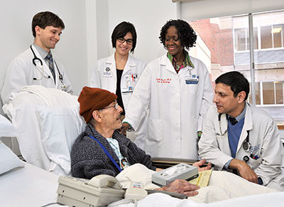
The team, whose meetings with patients are ongoing, consists of, from right, hospitalist Balal Ahmed, MBBS, Dr. Julietta Fiscella, and residents and students. Dr. Fiscella provides the team with mini reviews of staining, cellular structure, and other aspects of surgical pathology.
In some cases, patients were perplexed about how a systemic disorder could be associated with skin lesions. Their pathology reports gave descriptive diagnoses with no distinct diagnoses. Patient (or family) questions revolved around how the diagnosis was made, including processing and staining of pathology specimens, the nature of cells, types of cancer, cause of cancer and metastasis, certainty of diagnosis (and reasons for uncertainty), and family risk.
We have the following suggestions based on our experience with this pilot. First, communication between pathologists and patients is best conducted in conjunction with the primary clinician. Use of multidisciplinary teams facilitates integration of patient (and family member) questions about pathologic images with clinical implications. It allows the pathologist to discuss areas where she is expert while deferring clinical questions to the attending clinician. This approach maximizes transparency while minimizing the potential for miscommunication, including unintended blame.
Second, it is important for the pathologist to show empathy.7,10,11 Viewing a pathology image of cancer or other life-threatening diseases can have a profound impact on patients and their families. This requires skills that typically are not taught during pathology training and may not come easily to pathologists12,13—observing and responding to visual cues from the patient or pausing to allow the patient time to process the information and its emotional impact. A slow, deliberate, and compassionate approach goes a long way.
Third, it is important to explain pathology in lay terms.14 Medicine itself is specialized. Not infrequently, there is a lack of communication among the physicians themselves. Pathologists in particular are accustomed to using many pathological terms and most times these can be confusing to clinicians.15 The clinicians may find it difficult to explain to the patient what is meant in the pathology report, which adds another level of confusion for the patient.2 Using clear and simple language to explain diagnoses can be challenging but is critical.16–18 Words like “a small piece of your lung” can be substituted for “lung biopsy.” A phrase like “small balloon-like structures” can be substituted for “alveoli.”
While our pilot project was successful, it was not without challenges. Setting up a multidisciplinary conference at the patient bedside requires a scheduled time for the patients, families, primary clinician, resident, and pathologist. Making use of multidisciplinary teams in this way can be logistically difficult and time-intensive.19
We discovered that practicing clinical physicians have difficulty understanding and synthesizing the information in a pathology report. I provided the clinical team with mini review sessions on staining techniques, cellular structure, and other aspects of surgical pathology.
Each patient session lasted about 60 minutes. Additional time was required to prepare slides for the consultation. There is no system for reimbursing pathologists for performing this service. However, as health care moves from volume to value payments,20 this model may become economically viable.
In conclusion, our experience suggests that patients respond favorably to direct patient-pathologist-clinician communication. Given growing patient access to health information, including images, we encourage other pathologists to try our model.
References
- Blumenthal D, Tavenner M. The “meaningful use” regulation for electronic health records. N Engl J Med. 2010;363(6):501–504.
- Powsner SM, Costa J, Homer RJ. Clinicians are from Mars and pathologists are from Venus. Arch Pathol Lab Med. 2000; 124(7):1040–1046.
- Manek S. The pathology clinic—pathologists should see patients. Cytopathology. 2012;23(3):146–149.
- Dawson R, Gilbertson J, Kim S, Becich M. Pathology imaging on the Web. Extending the role of the pathologist as educator to patients. Clin Lab Med. 1999;19(4):849–866, vii.
- Epstein JI. The FAQ initiative explaining pathology reports to patients. Am J Surg Pathol. 2010;34(7):1058–1060.
- Wolfson WL. The FAQ initiative. Am J Surg Pathol. 2010;34(11):1728.
- Gutmann EJ. Pathologists and patients: can we talk? Mod Pathol. 2003;16(5):515–518.
- Gardner HA. The changing role of the pathologist. CMAJ. 1997;157(10):1346, 1348.
- Watt PC, Caughley L, Varma M. Should pathologists talk to patients? BMJ. 1990; 300(6731):1079.
- Zinn W. The empathic physician. Arch Intern Med. 1993;153(3):306–312.
- Buckman R, Tulsky JA, Rodin G. Empathic responses in clinical practice: intuition or tuition? CMAJ. 2011;183(5):569–571.
- Hung T, Jarvis-Selinger S, Ford JC. Residency choices by graduating medical students: why not pathology? Hum Pathol. 2011;42(6):802–807.
- Lambert TW, Goldacre MJ, Turner G, Domizio P, du Boulay C. Career choices for pathology: national surveys of graduates of 1974–2002 from UK medical schools. J Pathol. 2006;208(3):446–452.
- Research AfHQa. Health Literacy Universal Precautions Toolkit. 2011. www.ahrq.gov/qual/literacy, confirmed Nov. 6, 2011.
- Nakhleh RE. Quality in surgical pathology communication and reporting. Arch Pathol Lab Med. 2011;135(11):1394–1397.
- Houts PS, Doak CC, Doak LG, Loscalzo MJ. The role of pictures in improving health communication: a review of research on attention, comprehension, recall, and adherence. Patient Educ Couns. 2006;61(2):173–190.
- Doak CC, Doak LG, Friedell GH, Meade CD. Improving comprehension for cancer patients with low literacy skills: strategies for clinicians. CA Cancer Clin. 1998;48(3):151–162.
- Clear Health Communication. Pfizer Principles for Clear Health Communication: A Handbook for Creating Patient Education Materials That Enhance Understanding and Promote Health Outcomes. 2nd ed. www.pfizerhealthliteracy.com/asset/pdf/PfizerPrinciples.pdf.
- Kane B, Luz S, O’Briain DS, McDermott R. Multidisciplinary team meetings and their impact on workflow in radiology and pathology departments. BMC Med. 2007;5:15.
- Fisher ES, Shortell SM. Accountable care organizations: accountable for what, to whom, and how. JAMA. 2010;304(15): 1715–1716.
[hr]
Dr. Fiscella is chief, Department of Pathology, Cytopathology, and Laboratory Medicine, Strong Health Highland Hospital, Rochester, NY; clinical associate professor, pathology and laboratory medicine, University of Rochester Medical Center; and chair, Quality and Compliance Department, URMC Laboratory. Dr. Ahmed, the hospitalist who participated in the pilot, is associate chief of medicine, Strong Health Highland Hospital, and associate program director of the internal medicine residency program, University of Rochester.