NYU adapting VR technology to tell a pathology ‘story’
April 2019—The R&B classic “Time Is on My Side” may be an anthem for rejected lovers, but a new virtual reality teaching tool that allows students to “visit” the pathology lab without leaving the classroom may soon have NYU medical students humming the song’s refrain.
The experiential learning content, being developed collaboratively by multimedia specialists and pathologists, is designed to “maximize students’ time by condensing complex information,” says Greg Dorsainville, senior multimedia developer for NYU’s Institute for Innovations in Medical Education. Case in point: He took an hour of footage for the project’s pilot video, which shows a pathologist demonstrating a colorectal cancer staging. The final product clocks in at six minutes.
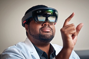
Greg Dorsainville, senior multimedia developer at NYU’s Institute for Innovations in Medical Education, experiments with virtual reality technology for pathology. (Photos courtesy of NYU Langone Health)
But the content isn’t merely intended as a timesaver for overstretched students. Dorsainville and Amy Rapkiewicz, MD, vice chair of pathology at NYU Winthrop Hospital, hope it will help teach pathology concepts—and how decisions made at the grossing table affect patient care—to medical students who have limited access to the pathology lab and no preclinical standalone courses in the specialty.
In the 1970s, says Dr. Rapkiewicz, who is also director of the integrated anatomy, histology, and pathology course at the NYU Long Island School of Medicine, medical schools began revising their preclinical curriculums to follow a systems-based approach in which traditional sciences like pathology are taught as a component of courses on the organ systems. “This was spurred on by the finding that students seemed to do better contextualizing the basic sciences when they’re learning about clinical sciences at the same time,” she explains. Consequently, educators have had to determine not only what to teach students in their preclinical years but how to teach it to them.
This led to a eureka moment of sorts that developed organically from a long collaborative partnership between Dorsainville and Dr. Rapkiewicz, who initially worked together to create a digital catalog of pathology specimens. The two moved away from that project, Dr. Rapkiewicz says, after realizing students were unlikely to benefit from the library without a structured didactic approach.
But Dr. Rapkiewicz, a forensic pathologist and autopsy director, had repeatedly seen material click for students who visited the autopsy suite and interacted directly with specimens. When she shared this insight with Dorsainville, he suggested immersive content, giving students “a feeling of presence, as if they’re standing next to the professor in the lab.”
Dorsainville, an experienced videographer who’s been working with immersive technologies for three years, uses a 360-degree wide-angle camera to film the video content. The camera, which captures a much larger field of vision than a traditional video camera, “gets as much of the view as possible but also gives a stereo feel,” he says.
The videos, which can be viewed from a virtual reality headset or a desktop computer, aren’t difficult to develop from a technical standpoint. What’s tricky, Dorsainville says, is “creating a viable educational experience.”
The example video of the colorectal cancer staging is in usability testing with a group of preclinical students at the NYU School of Medicine. All content will be pilot tested before becoming part of the curriculum. Because crafting an effective pedagogical strategy is challenging, Dr. Rapkiewicz is working with colleague Margret Magid, MD, pathology content director, on how best to implement the videos in integrated coursework.
In the meantime, the development team is focused on creating content that has what Dr. Rapkiewicz calls “a robust pathology story.” A post-chemotherapeutic osteosarcoma, for example, has a clean narrative with an illustrative surgical specimen. A lesson featuring an osteosarcoma might show the x-ray, chemotherapeutic effect, and surgical amputation, culminating with the oncologic targets and revealing the extensive role pathologists play in a patient’s downstream clinical management. A sarcoma of the soft tissue, which “kind of looks like a blob,” would not be a good candidate, she notes.

Dr. Rapkiewicz
“A lot of times I see people make wonderful simulations or innovative VR content and it’s something I may be able to explain to students [by drawing] on a napkin,” Dr. Rapkiewicz says with a laugh. “We’re really trying to use it to fit the technology with the problem.”
For preclinical instruction, this means using the content to augment areas of the curriculum for which students might need “extra context to understand how pathology and disease are represented in the body,” Dr. Rapkiewicz says. For clinical instruction, she envisions directing students to supplementary pathology material—for example, students on a surgery rotation might be given a list of immersive videos to view after observing a gallbladder surgery. This “just-in-time learning” will be essential for students who don’t have time to “follow their specimen to the pathology lab,” she adds.
The first project of Dorsainville and Dr. Rapkiewicz to be incorporated into the preclinical curriculum is tangential to pathology. Beginning this fall, students in the general anatomy course at the NYU Long Island School of Medicine will use virtual prosection modules, in lieu of cadavers, in the introductory anatomy course. “Our strategy right now is to follow responsive design—for all this content to be accessible in some format, regardless of whether students have a headset, computer, or mobile device,” explains Dorsainville. “All experiences will be available via the Web. For true immersion, students will have access to devices loaned by the school.”
Dorsainville, meanwhile, hopes to further develop other virtual reality pathology content by adding an interactive component that allows students to manipulate digital models of the tissue, similar to what he’s done with the prosection modules.
“The only thing really lacking,” he says, “is that you can’t walk around and pick things up.” —Charna Albert
Xifin adds functionality to LIS and RCM system
Xifin has announced a strategic partnership with Glidian under which Glidian will integrate its automated prior authorization capabilities with the Xifin RPM revenue cycle management solution and Xifin’s laboratory information system.
“By integrating our electronic prior authorization submissions with Xifin RPM and Xifin LIS, we’re able to help providers streamline their workflows, prevent denials, and help expedite decisions to make sure patients receive the important diagnostic information they need,” said Ashish Dua, CEO of Glidian, in a press statement. The application works with the existing prior authorization workflow, logic, and document storage in Xifin RPM.
Xifin has also introduced a patient responsibility estimator for Xifin RPM, which is designed to enhance patients’ understanding of out-of-pocket costs for ordered tests. The estimator, which is available to physicians and patients through Xifin’s multi-portal infrastructure, also offers patients a prepayment option.
Xifin, 866-934-6364
Paige.AI receives FDA breakthrough designation
The FDA has awarded Paige.AI breakthrough device designation for its use of computational pathology to build artificial intelligence tools for cancer diagnosis and treatment.
“We are thrilled to receive breakthrough designation and look forward to collaborating with the FDA to bring our products to market, starting with prostate cancer and expanding from there,” said Leo Grady, PhD, CEO of Paige.AI, in a company press release.
Paige.AI has received more than 1 million deidentified images of digitized slides from Memorial Sloan Kettering Cancer Center under a license agreement with that institution. The company is funding the digitization of an additional 4 million slides from MSK’s archives to develop a portfolio of artificial intelligence products across cancer subtypes for use by pathologists.
The FDA grants breakthrough device designation for technologies that have the potential to provide more effective diagnosis or treatment for life-threatening or irreversibly debilitating diseases or conditions than alternative approved products or where no alternative exists.
Northwest Pathology implements LigoLab billing/RCM system
LigoLab Information Systems has announced that Northwest Pathology, Bellingham, Wash., recently began using its comprehensive billing/revenue cycle management solution.
The billing/RCM module is provided as part of the LigoLab integrated information system, which also includes anatomic and clinical pathology and molecular diagnostics.
The billing/RCM system offers real-time patient demographic checking and patient insurance eligibility options, automatic CPT and diagnosis coding, claim scrubbing, advance beneficiary notice generation, and patient and client billing. It includes a screen for patient encounters that displays the billing record and claim status, corresponding codes and charges, scans of associated case documents, and reports.
LigoLab, 800-544-6522
Cybersecurity toolkit for small to midsize companies
The Global Cyber Alliance and Mastercard have released a free online cybersecurity toolkit for small and medium-sized businesses.
The toolkit helps users take inventory of cyber-related assets, create and maintain strong passwords, use multi-factor authentication, back up critical data, and prevent phishing and viruses. It provides templates of policies and forms, training videos, and customizable documents.
“What sets the Global Cyber Alliance Cybersecurity Toolkit apart is that it is an action kit,” said Philip Reitinger, president and CEO of the Global Cyber Alliance, in a press statement. “Our focus is on producing a dynamic clearinghouse of operational tools that help small and medium [sized] businesses address risk and improve their cybersecurity posture, leveraging the deep expertise of our network of global partners, such as Mastercard, and the experiences of actual GCA toolkit users.”
The Global Cyber Alliance partnered with several organizations to develop the toolkit, including the Center for Internet Security, Cyber Readiness Institute, and cities of London and New York. The guide will be updated regularly with input from users, industry experts, and public and private partners worldwide.
The toolkit is available at https://gcatoolkit.org/smallbusiness.
Cleveland Clinic launches AI innovation center
The Cleveland Clinic has established the Center for Clinical Artificial Intelligence to develop clinical applications for AI.
The innovation hub will bring together specialists from several departments, including pathology, imaging, information technology, oncology, genomics, and quantitative health sciences.
“The center will serve as a platform for collaboration and communication between physicians and data scientists; provide programmatic and technology support for AI initiatives; and conduct research in several areas of medicine that can solve clinical problems using machine learning, deep learning, and other AI technologies,” according to an announcement from the Cleveland Clinic.
The health care institution expects the center, a project of Cleveland Clinic Enterprise Analytics, to have global reach and include collaborations between industry and academia.
Already underway are projects to build machine learning models that identify patients who have a high risk of death within 48 to 72 hours of admission and predict inpatient length of stay and readmission risk. Researchers are also undertaking cancer-focused projects to predict patient outcomes.
Dr. Aller teaches informatics in the Department of Pathology, University of Southern California, Los Angeles. He can be reached at raller@usc.edu. Hal Weiner is president of Weiner Consulting Services LLC, Eugene, Ore. He can be reached at hal@weinerconsulting.com.