Editor: Frederick L. Kiechle, MD, PhD
Submit your pathology-related question for reply by appropriate medical consultants. CAP TODAY will make every effort to answer all relevant questions. However, those questions that are not of general interest may not receive a reply. For your question to be considered, you must include your name and address; this information will be omitted if your question is published in CAP TODAY.
Q. What are the guidelines for proper handling and processing of blood specimens collected in serum separator tubes?
A. Following blood collection, serum separator tubes (SST) should be gently inverted by five complete inversions to disperse the clot activator—the silica particles that coat the walls of the tubes—into the blood. Note that one inversion is turning the tube upside down and returning it to its upright position. The specimen should be thoroughly mixed, as inadequate mixing may result in incomplete clotting and the formation of fibrin; this may interfere with barrier formation. Avoid vigorous mixing or shaking of the specimen because this may cause hemolysis, which may require redraw.1
SST tubes should be allowed to clot for 30 minutes in a vertical position in a tube rack; a dense clot should be observed. Specimens from certain patient populations with impaired coagulation may require longer than 30 minutes to clot in SST tubes. These include patients on anticoagulant therapy, on high doses of heparin (no clotting may occur), and with certain diseases (e.g. liver disease or multiple myeloma).
It is recommended that serum be physically separated from contact with cells by centrifugation as soon as possible with a maximum time limit of two hours from the time of collection, unless there is conclusive evidence that longer contact times do not contribute to error in the results.
SST tubes should not be recentrifuged once the barrier has formed, as this may increase the concentration of certain analytes (i.e. glucose, sodium, potassium, creatinine, phosphorus).2 If recentrifugation is required for improved serum quality, serum above the gel should be aspirated into a properly labeled, clean tube, which then can be centrifuged.
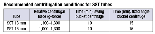 These recommendations are for the BD Vacutainer SST tube. Laboratory professionals should consult manufacturer recommendations. For additional information, visit www.bd.com/vacutainer/techtalk (“The importance of properly processing a BD Vacutainer SST tube”) or www.specimencare.com.
- Milutinović D, Andrijević I, Ličina M, Andrijević L. Confidence level in venipuncture and knowledge on causes of in vitro hemolysis among healthcare professionals. Biochem Med. 2015;25(3):401–409.
- Shafi H, Sadrzadeh H. The effect of recentrifugation of serum separator tubes on concentration of serum analytes. Ann Clin Lab Sci. 2012;42(3):318–319.
Sol F. Green, PhD, Director of Medical Affairs, Clinical Process Optimization, and Global Technical Services, BD Life Sciences, Franklin Lakes, NJ, Assistant Professor of Clinical Pathology, Stony Brook School of Medicine, NY
Lena Arzoumanian, Senior Technical Services Specialist, BD Life Sciences, Franklin Lakes, NJ
[hr]
Q. Are there regulations guiding the practice of taking additional blood samples from a patient even though there are no orders for the blood samples? These “just in case” specimens are sent to our laboratory by the emergency department when a port or catheter is placed in the patient. The ED’s reasoning is that it prevents a patient from being stuck twice if there is an order for blood tests later. Our lab has to either store the samples or process them (centrifuge or separate RBCs from serum) so they are ready in case an order is entered later. Should this practice be banned? Should we refuse to accept these samples?
A. I do not believe there are regulations prohibiting this practice; however, the “in case of” and the “collect a rainbow” practices are not, in my opinion, laboratory medicine best practices, as both represent the tip of the inpatient inappropriate laboratory test utilization iceberg that affects all of us.
As laboratorians we need to have zero tolerance for any unnecessary phlebotomy because those incremental 5 cc’s add up very quickly. The limited literature on this topic has shown that inpatients (in those studies) have had up to 700 mL of blood collected during hospital stays. We have to step back and ask: Does that make sense? Does it make sense to collect an extra tube or tubes just because there might be something else to test? We need to look at these practices and determine the following:
- How often have we used the extra tube?
- How many tubes of the rainbow have we not used?
- How often do we even do “add on” tests?
- We then need to review our inpatients and determine how often comprehensive and basic metabolic panels, etc., are being drawn per stay, per day, and then review serial results to determine if there was a change in lab results from collection to collection.
- We then need to calculate how much blood was collected (3–5 mL per PST/SST, 2–4 mL per CBC, and 3–5 mL per coagulation study); the results will be scary.
I’m sure readers will not be surprised by how much blood is being wasted at their individual institutions. At my institution, we have gone from the “rainbow” to the extra tube to no extra tube “except as driven by clinician request.” We have done the “add on” study as well and found that very few (10 percent) tests were added on after day two.
The common complaint from clinical patient care providers is “just use smaller tubes.” However, the problem is not the size of the tube but the number of tubes—that is, too much potentially unnecessary laboratory testing. Let’s reduce the latter and then fix the former.
- Wisser D, van Ackern K, Knoll E, Wisser H, Bertsch T. Blood loss from laboratory tests. Clin Chem. 2003;49(10):1651–1655.
- Hicks JM. Excessive blood drawing for laboratory tests. N Engl J Med. 1999;340(21):1690.
- Foulke GE, Harlow DJ. Effective measures for reducing blood loss from diagnostic laboratory tests in intensive care patients. Crit Care Med. 1989;17(11):1143–1145.
- Smoller BR, Kruskall MS. Phlebotomy for diagnostic laboratory tests in adults. Pattern of use and effect on transfusion requirements. N Engl J Med. 1986;314(19):1233–1235.
- Henry ML, Garner WL, Fabri PJ. Iatrogenic anemia. Am J Surg. 1986;151(3):362–363.
David N. Alter, MD, DABCC, Clinical/Chemical Pathologist, Point of Care Testing–System Medical Director, Spectrum Health Regional Laboratory, Grand Rapids, Mich.
Clinical Professor of Pathology, College of Human Medicine, Michigan State University, Vice Chair, CAP Chemistry, Resource Committee
[hr]
Dr. Kiechle is medical director of clinical pathology, Memorial Healthcare, Hollywood, Fla. Use the reader service card to submit your inquiries, or address them to Sherrie Rice, CAP TODAY, 325 Waukegan Road, Northfield, IL 60093; srice@cap.org. Those questions that are of general interest will be answered.