Karen Titus
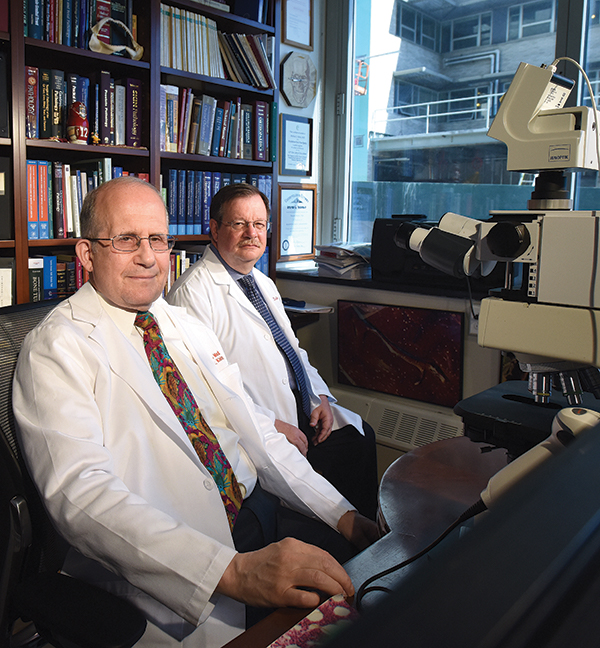
Dr. Michael Klein (left) and Dr. Edward DiCarlo
made a compelling case in 2014 for comparing clinical and histologic diagnoses in patients
undergoing total joint arthroplasties.
July 2017—One of the more unnerving scenes in contemporary theater comes courtesy of Martin McDonagh’s “A Skull in Connemara,” which opens with two men in an Irish graveyard, hired by the local priest to make room in the overcrowded burial ground. Their method? Exhume the corpses and smash the bones to bits.
The action is macabre and outrageous. Practically speaking, it’s also efficient (and in McDonagh’s hands, distressingly funny). What else can one do with bones that don’t seem to matter, that are old and now in the way?
A similar question taunts pathologists and surgeons. When performing total joint arthroplasties, what should surgeons do with the hip and knee specimens? Submit them for pathologic examination? If so, is a gross examination sufficient? When might a microscopic examination be in order? Is it ever okay to channel McDonagh’s Irishmen and toss the bones aside as (biohazardous) rubbish?
A look at the orthopedic literature suggests the latter approach might actually have fiscal soundness. The message is clear from titles such as: “Histologic examinations of arthroplasty specimens are not cost-effective” (Lin MM, et al. Clin Orthop Relat Res. 2012;470[5]:1452–1460). The point of these studies, generally, is that routine pathologic exams boost costs but rarely alter patient management. The phrases “limited cost-effectiveness” and “low prevalence of findings” pop up with the regularity of a president on a golf course.
The reception these studies receive from pathologists, however, can be chilly.
Take, for example, a letter spearheaded by James Richard, DO, who responded to a study, which took place at the institutions where he practices, that suggested histologic examinations of shoulder arthroscopies did not influence patient care or provide new diagnoses. In the letter, published in the Journal of Bone & Joint Surgery in November 2008, Dr. Richard, who is director of laboratories, Sparrow Health System, Lansing, Mich., argued the value of doing such exams. In one 12-month period alone, he and his colleagues wrote, they uncovered nine cases of malignancy or probable malignancy from the examination of all orthopedic specimens (not just shoulder arthroscopies).
Dr. Richard’s views remain undimmed nearly a decade later, in 2017. Sparrow does not routinely do microscopic exams. “We’re not even receiving the specimens,” he says.
Dr. Richard says the decision was made before his arrival. Bringing it up for discussion “would be opening Pandora’s box,” he says. At another institution where histologic exams were routine, he had to defend the practice on more than one occasion. He succeeded, but since his departure, he notes, the institution has stopped sending total joint specimens to pathology. He sounds a bit weary as he weighs his efforts over the years. “I’ve been tilting at that windmill for quite some time.”
Keep tilting, says Michael Klein, MD. With a 16,587-specimen study to back up his points (DiCarlo EF, Klein MJ. Am J Clin Pathol. 2014;141[1]:111–118), Dr. Klein makes a compelling case for comparing clinical and histologic diagnoses. As he points out, most previous studies top out at 1,200 or 1,500 cases. “And those series are usually collected from a couple of hospitals and combined,” says Dr. Klein, pathologist-in-chief emeritus, Department of Pathology and Laboratory Medicine, Hospital for Special Surgery, New York.
Dr. Klein, who is also a professor of pathology and laboratory medicine, Weill Cornell Medical College, and consultant in pathology, Memorial Sloan Kettering Cancer Center, sees more than his share of total arthroplasty specimens. At Special Surgery, there may be 40 to 60 total joint replacements a day. The topic is inherently relevant to his work, but he says his interest has also been provoked by the extensive orthopedic literature that suggests certain pathology—and even certain radiology and indeed surgical—procedures are not considered to be cost-effective and thus should not be done.
“It all sounds very altruistic,” he says. “It’s not exactly altruistic. I believe a little bit of it is self-serving and self-preserving, which I understand.” Moreover, he says, the orthopedic literature often neglects to point out reasons other than cost-effectiveness for doing these exams, including quality assurance, risk management, and, of course, patient care.
Dr. Klein also bristles at study authors who draw their conclusions based only on whether histologic exams identify a clinically unsuspected tumor. This is a fallacy, he says, since tumors are relatively rare anyway, and a careful gross examination is needed before a pathologist decides to take sections. A microscopic examination can identify tumor type, but it’s not needed to find the tumor to begin with.
Spurred by what he saw as the lack of useful studies, Dr. Klein and Edward DiCarlo, MD, undertook their own study, assessing total joint replacement specimens (7,968 hips, 8,619 knees) over a 10-year period that had been grossly and microscopically examined by Dr. DiCarlo (chief of surgical pathology at HSS) and verified by Dr. Klein.
Notably absent from their study was an attempt to provide cost analysis. The pathologists simply wanted to compare the postoperative surgical diagnosis with the pathologic diagnosis for the seven most common diagnoses: degenerative joint disease, traumatic injury/fracture, avascular necrosis, subchondral insufficiency fracture, rapidly progressive arthritis, inflammatory arthritis, and septic arthritis. Dr. Klein says they expected their study to mirror earlier orthopedic studies, which found no significant differences between clinical and histologic diagnoses.
The numbers told a much different story. The discrepancy rate was 18.8 percent for hips and 9.4 percent for knees. And 5.4 percent of hip joints and 1.4 percent of knee joints showed discordant histologic findings that were clinically unsuspected and should have affected patient management and outcomes.
Surgeons who say examinations aren’t needed because their clinical assessments are correct are, well, wrong, says Dr. Klein. There are, he says, important diagnostic findings apart from tumors, particularly subarticular insufficiency fractures. This diagnostic category is often mistaken for degenerative joint disease, but should be treated differently. “In fact, because its clinical history is different from degenerative joint disease, it should probably be diagnosed preoperatively by surgeons, and it’s not,” he says. In some cases, a total joint replacement may not be merited as a primary treatment. In fact, this condition is often associated with underlying comorbidities, such as morbid obesity, metabolic bone diseases, and lifestyle choices that may be altered with appropriate medical advice and treatment to prevent future fractures in other joints.
Degenerative joint disease was the most common diagnosis among surgeons and pathologists, but familiarity didn’t necessarily breed accuracy. It was also the most overdiagnosed condition—it was diagnosed approximately 20 percent more frequently than could be verified by the pathologists. And, the authors say, despite its prevalence, it was not always recognized clinically when it was present.

Dr. Bachner
Paul Bachner, MD, sings the praises of the Hospital for Special Surgery, noting its status as a highly acclaimed orthopedic hospital. “You have to assume their orthopedists are extremely experienced.” Nonetheless, the study uncovered those 18.8 and 9.4 percent error rates. “And this is a group that’s probably as expert as you can find in the country,” says Dr. Bachner, a professor of pathology and past chair of the Department of Pathology and Laboratory Medicine, University of Kentucky, Lexington.
(The flip side, he says, is that Drs. Klein and DiCarlo are two of the best orthopedic pathologists in the country. “Their ability to find things may be greater than a pathologist who does not specialize in that area.” As they themselves noted in their study, the two had an aggregate experience in bone disease analysis of more than 65 years.)
In his own experience, Dr. Bachner, who in June retired as director of laboratories at UK, says the most common finding is avascular necrosis in cases where there’s a radiologic and clinical diagnosis of osteoarthritis. “And every once in a while you’ll find a cancer—it’s very rare.”
Dr. Klein has plenty to say about histologic exams of total joint arthroplasties. But he’s succinct on one point in particular: Don’t abandon histologic exams because of so-called cost-effectiveness. “Cost-effectiveness isn’t the reason we do pathology.”
Another reason to do histologic examinations, Dr. Klein says, is to learn what the actual disease rate is in a series of surgeries on a site for a particular surgeon, just as one would for any organ system. Before the advent of CT scanning, the acceptable excision rate of normal appendices for clinically suspected acute appendicitis was 15 percent. Significantly more than this and a surgeon was too aggressive; significantly less would mean the surgeon was too conservative. Is there an acceptable rate for removing histologically normal joints? “While this ideally should not happen, there is probably some very small finite percentage that is permissible,” Dr. Klein says. He has no idea what that number is, but determining the rate is important. “If you throw that information out, then you’re neglecting an essential part of public health.”
Dr. Richard links the matter to the broader picture as well. He’s familiar with the arguments that a bone might have broken based on circumstances—say, an auto accident. “But was there anything causing a weakening for it to be broken at that specific site, that specific bone? Was there an unknown defect that caused it?” That’s just as true for hip joints, he says. “How many times do we hear in the literature about patients falling and fracturing a hip?” Pathology might reveal an occult malignancy; it might also reveal metastatic spread. If the pathologist finds crystals, it may be indicative of a systemic disease. “There’ve been times when we’ve picked up acute inflammation” indicating possible sepsis. “And there are times when you find what might be considered subtle changes, but they aren’t—they’re diagnostic.” The obvious, in short, isn’t always obvious. “It goes back to the diagnosis and documentation that are part of our role.”
Dr. Richard urges pathologists to take what he calls the higher road: concern about patient care and patient safety. “That’s where we’ve got to hang our hat.” At the minimum, pathologists should look at all fracture cases, he says, or any cases the surgeon deems unusual.
“We’re guardians of the galaxy here, if you will. We’re trying to prevent potential threats, hoping they never come to fruition.” — James Richard, DO
“I would encourage, for documentation purposes, all specimens be submitted,” he continues, “but a majority of them would be for gross-only evaluation.” Microscopic evaluations would occur only if the pathologist thought the case was unusual enough to merit a closer look, or if the surgeon requested it, based on the patient’s history and clinical information. “Most of us in pathology recognize standard degenerative changes within that joint surface,” Dr. Richard says. “We see it, we document it, we put out a simple report saying, ‘Yes, it is consistent with that.’ And that would be the end of it.”
Such efforts are, he says, the equivalent of following the rules of the road. A driver at an empty intersection stops at the stop sign, he says, “even if it’s in the middle of the Utah desert and you can’t see anyone for miles in any direction. This is what you do.”
So why might some physicians be tempted to blow through that stop sign? Perhaps the answer has something to do with the dollar sign.
Dr. Klein suspects that if payment is perceived to be at the heart of the matter, pathologists will find it hard to bring up the issue. “Because why all of a sudden are we so interested?” he asks. Surgeons might assume it’s because pathologists want to collect a professional component. “Why else would we care?”
In the case of bundled payments, money for the pathology work comes out of—to put it crudely—the surgeon’s pocket. Although the pathology payment might be small, some surgeons refuse to part with it, says a pathologist whose pleas to surgeons and administrators to allow histologic exams of total joint fell on deaf ears. “No one wanted to give up any portion—in this case, $100—of the bundled payments to the lab. So now the surgeons just throw the hips away.”
Pathologists have financial incentives, too, of course. In smaller practices especially, Dr. Richard says, “Every little case is important financially.”
If pathologists are questioned about their financial incentives, Dr. Richard has a simple suggestion: Offer to cut your fee by 10 percent for a gross-only exam if the surgeon will cut their fee by the same percent. That should put a quick end to the discussion, he jokes, and bring the focus back where it belongs.
“We should look at everything that comes out
of the patient. It’s our job. ” — Nicole Riddle, MD
Even without slashing fees, examining hips is not a money-printing enterprise, points out Nicole D. Riddle, MD, staff pathologist, Tampa (Fla.) General Hospital. With a billing code of 88304, hips, in and of themselves, are on par with appendices, gallbladders, and benign skin cysts, she says. “It’s not that we’re trying to churn out hips to get rich,” says Dr. Riddle, who is also an assistant professor of pathology, University of South Florida, Tampa.
Dr. Bachner can attest to that. Several years earlier, some of his surgical colleagues proposed changing the policy. At that time, Dr. Bachner used data from Dr. Klein’s study to bolster his position. He also did two surveys of his own, sending questionnaires to members of both the Association of Pathology Chairs and the Association of Directors of Anatomic and Surgical Pathology. Of the 46 responses, about 80 percent said they did routine histologic examination. Of the personal comments he received, Dr. Bachner says most were from those who felt it was beneficial both clinically and for QA. But when he summarized his findings for his colleagues, he tried to construct a pie chart to illustrate the financial implications. The software wouldn’t accept the number he plugged in for the cost of pathology. “It was so small,” he explains.
Dr. Klein recalls an incident at another institution when a senior resident asked him about findings on a femoral head—osteoarthritis, as it turned out. After he explained the pathology in detail, his colleague said this was her first encounter with such a case. Dr. Klein learned, much to his surprise, that all femoral heads as well as knees were being thrown out. “It practically took an act of Congress to get the surgeons to begin sending that stuff,” he says. Change came only when a new physician in chief was appointed at the institution. Dr. Klein approached her only after ascertaining that he had the support of his department chair. When Dr. Klein explained the QA and medical-legal implications, the practice changed. “Of course, what made the real difference in getting the support of the chair was explaining that $400,000 a year of potential professional billings were getting thrown out,” he says. “I got the backing, but it wasn’t because he was interested in femoral heads.”
In practices where money is less of an issue, Dr. Richard says, so-called little cases might seem like more trouble than they’re worth. “They might possibly say, ‘Oh, the last thing I want to do is look at 20 slides of arthroscopy shavings.’ ”
Dr. Bachner dittos that point. Though he successfully lobbied to retain the practice at UK a few years back, more recently his own department decided to forego routine histopathologic examinations of all total joint arthroplasties. “Much to my chagrin,” he says. Instead, they put together a series of indications on gross examinations for performing microscopic exams on hips and knees. “Time will tell how that works out,” says Dr. Bachner. His colleagues are busy, he notes, and are looking for ways to concentrate their efforts in areas they deem more relevant than total joint exams. He speaks politely and chooses his words carefully.
When he speaks of the topic more generally, his personal—“And I stress the word ‘personal’ ”—feeling is less circumspect. It’s easy to question the usefulness of longtime medical practices, he says, and once you question and discontinue one practice, it only becomes easier to jettison other tasks. “This represents another move toward the dumbing down of medicine,” says Dr. Bachner.
Asked by CAP TODAY about responses to the paper, Dr. Klein jokes about turning off the reporter’s recording device first. He then plunges ahead with a lengthy narrative: how they submitted it to several nonpathology publications first and were met with both disdain and enthusiasm; how he feared the results might reflect badly on the surgical colleagues he highly respects; how he battled what he considered to be prejudicial peer review from one journal.
Such perils aren’t exclusive to publishing. Pathologists are careful not to point accusatory fingers at their colleagues either inside or outside the lab. But a certain frustration creeps into many conversations. As in the 2016 presidential election, when many voters struggled with how, or if, to address complicated and incendiary issues of gender, race, religion, and class, pathologists sometimes find themselves wondering how to talk about the total joint specimens. How best to ask important but possibly sensitive questions?
The histopathologic exam serves as a check on a surgeon’s work, but as Dr. Richard points out, not all orthopedic surgeons welcome having a pathologist peering over their shoulder.
Many do, of course. Philip Branton, MD, consultant, Biorepositories and Biospecimen Research Branch, National Cancer Institute, and chair of the CAP’s Biorepository Accreditation Program advisory group, says he’s never worked at an institution where an orthopedic surgeon has questioned the practice of joint examinations. “Maybe I was just incredibly lucky and worked with unique sets of clinicians,” he muses. He recalls having exactly zero arguments with clinicians about specimen evaluation. Surgeons’ attitudes were more one of relief, he says: Once it leaves the OR, it’s your guys’ problem, went the thinking.
Reactions can vary with specialty. With obstetrician-gynecologists, says Dr. Richard, “There isn’t a specimen they take out that doesn’t go to pathology.” Even Fallopian tubes that appear completely normal will make their way to pathology, since surgeons are concerned about small changes that may not be evident to them.
The current CAP policy leaves pathologists and medical staff to decide locally which surgical specimens to submit and which to exempt. Dr. Richard, who is speaker of the CAP House of Delegates, says this remains the best approach. But in calling for physicians at each institution to decide as a group, pathologists may feel they’re at a disadvantage, he acknowledges. Who has the bigger department, surgeons or pathologists? Who brings in the most money? Who has the administration’s ear? “If the orthopod says he doesn’t want to do something, and there are 50 orthopods and 10 pathologists, I’ll be honest, that’s not one you’re going to take to the administration—unless you say, ‘I need your help because your insurance policy is at risk,’ ” says Dr. Richard.
Even in less fraught circumstances, surgeons might push back: What’s the benefit compared to the time I have? Tell me what problem you’re solving. Tell me what you’re helping me with if I say yes.
Dr. Richard spins out such a scenario with the detail and inflections of someone who’s heard it all before. It’s almost like a tiny theatrical performance. To wit:
Pathologist: We do this for everybody, for patient safety. If something comes out of the body, it comes through our department. And there’s a risk-management element.
Surgeon: I’ve never been sued for any of my arthroscopic surgeries.
Pathologist: Wonderful. That’s fantastic. All the same, if you miss a septic joint, what’s your risk on that?
Surgeon: Well, nobody’s ever sued me.
Pathologist: No, but the discussion might be different if some of your patients had known you might have identified it earlier.
Surgeon: You see a thousand arthroscopies and you might only find one.
Pathologist: That’s a valid point. Let’s turn it around—how long is that patient septic if the diagnosis is delayed? What’s the greatest risk?
Dr. Riddle has her own riff (also complete with voices) on such conversations. She suspects that some surgeons who say exams are unnecessary because findings are rare “are smart enough to realize they shouldn’t say they don’t want to give money to pathology.” But others are less careful with their words. “I’ve had people say, ‘It’s X amount of dollars—why should I?’ Or, they tie both rationales together: ‘It’s X amount. Why should I when they rarely find anything?’ ”
She also takes aim at the circular reasoning used to say findings are rare. How do you know if you don’t look? she asks. Specimens are routinely submitted for examination at Tampa General Hospital, and she and her colleagues have found polyomavirus, hematopoietic malignancies, and metastatic tumors, all previously unknown.
Are surgeons happy these are caught? Dr. Riddle pauses for an interesting interval. “Usually.” Pause. “Of course. They want what’s best for the patient.” But, she says, some personalities deal better with mistakes than others. And some of those others “get upset when we find something and surprise them with it.”
That may be human nature, but Dr. Richard urges pathologists to keep the larger picture in mind. “We’re guardians of the galaxy here, if you will,” says Dr. Richard. “We’re trying to prevent potential threats, hoping they never come to fruition.” The biggest mistake pathologists make in these discussions, he says, is making it a personal issue. Avoid these useless detours, he says. “It needs to become a matter of having a serious discussion that takes finances out of it and goes directly to patient care and safety.” Put another way, if you want to drive from San Francisco to Los Angeles, follow the coast—there’s no need to hit Fresno or Bakersfield.
Focus on the data, Dr. Bachner says, starting with the DiCarlo/Klein paper. “It’s clearly the best paper in the field. There’s nothing even near it in terms of breadth and scope.”
Dr. Branton
Dr. Branton, who is past chair of the CAP Surgical Pathology Committee, weighs in with a cautionary thought. “My personal philosophy would be that if you don’t examine specimens, you’re doing so not at your own peril but at your patient’s peril. Sooner or later something is going to be missed.”
When the what-if talk subsides, the matter of actually doing histologic exams can seem almost like an afterthought. How difficult is the task?
Dr. Klein’s place of practice gives him an unusual take on matters. Special Surgery, an all-orthopedic institution, handles 20,000-some orthopedic—soft tissue or bone—specimens a year, nearly all of which undergo a microscopic examination. A defined institutional exclusion list eliminates a few specimens, such as tiny osteophyte bunions. (“Even so, a few surgeons want them done,” Dr. Klein says.) Otherwise, pathologists will take that closer look. “If they revise a prosthesis and there’s tissue attached to [it], we evaluate that tissue. We want to see if there’s metal or cement. We want to see if there’s possibly an infection. If there’s particle disease, we want to see the size and type of the particles because we want to give them some idea of why the prosthesis wore out. There’s a whole variety of things to evaluate.”
Is this a realistic approach for pathologists at less-specialized institutions? Dr. Klein thinks so; the big reason they don’t, he says, “is lack of interest.” That’s followed by technical and clerical challenges. “It takes motivation to decalcify tissues,” he says.
Those who are motivated may not always be acting efficiently, he suggests. When he consults on total joints removed from outside cases, he says he invariably sees five to seven slides, sometimes none of which demonstrate the area described grossly as having degenerative disease. “And yet they’ve made half a dozen histologic slides of material that’s been decalcified.”
“In our institution we make one decalcified section per case. One,” Dr. Klein says. “That’s the routine. Very exceptionally we’ll do two or three, but that’s only if a patient has ochronosis or some incredibly interesting pathologic condition.” But to document a degenerative disease or just about any other kind of joint pathology, only two sections are needed, says Dr. Klein, one from the diseased surface and one synovial tissue, to pick up inflammatory joint diseases or other undiagnosed conditions.
“You just need to document the right places,” Dr. Klein continues. “With a total knee, you can take two or three bits of hard tissue and put them in one decalcified cassette, and you have the entire story.” He also suggests that the decalcification process can be streamlined “if people are motivated enough. You can literally do the fixation and decalcification at the same time if you use the right solution.” While such solutions are generally not commercially available and must be made by the pathologist or technologist, “They shorten time from receipt of tissue to diagnosis by two days.”
Then there’s the issue of carpentry. To make the sections, “How do I saw them?” Dr. Klein asks. “Do I use a band saw, which is dangerous?” It may not make sense to invest in expensive, specialized equipment unless the institution sees a fair amount of osseous specimens, he says. Otherwise, a safe, albeit arm-tiring, double-bladed hacksaw is the best option. “We used to cut all our gross specimens with a butcher band saw. That was dangerous. You could cut your fingers off. I know bone pathologists who’ve injured themselves on those blades.” Currently he and his colleagues use a saw with no teeth, a metallic blade with a diamond carbide edge. “It’s an expensive saw, but it won’t cut you. You literally have to press your hand against it while it’s running full blast, and then it will only nick your skin.”
What about places with lower volumes? “It’s my opinion that all total joints should be sectioned,” Dr. Klein says. “Whether they should be sectioned by someone in a small community hospital, that’s another issue.” It might be worth considering sending samples to regional centers. “Personally, I think that’s the way to go. I think it’s much safer for patients and for public health data.”
Dr. Riddle takes a more sanguine view. “Any surgical pathologist working anywhere in the United States should have the training and capability to do this.” Indeed, she sounds like a DIYer on a home improvement project: “All you need is a handsaw.”
Dr. Bachner falls somewhere in the middle. Many pathologists are uncomfortable with orthopedic pathology, he says. Pathologists tend to experience it in two ways: hip and knee resections (“They find this somewhat of a chore—it’s a nuisance,” he says) and bone tumors (“Most are afraid of bone tumors, because they’re rare and they don’t have a lot of experience with them”).
Yet there’s plenty to be gained, it would appear, from maintaining or developing the practice of examining total joint arthroplasties.
Dr. Branton could not state matters more simply. “I think throwing bones in the trash is a bad idea.” (Sorry, Martin McDonagh.) Certainly it could arouse the curiosity of a malpractice lawyer. Joint examination is good, conservative medical practice, he says.
“We should look at everything that comes out of the patient,” Dr. Riddle agrees. “It’s our job.” Knocking the proverbial fat lady aside, she adds, “Nothing is final until pathology looks at it.”
[hr]
Karen Titus is CAP TODAY contributing editor and co-managing editor.