
Kaitlin E. Sundling, MD, PhD
Adele Kraft, MD
January 2021—Although the challenges we face due to the COVID-19 pandemic are significant, adapting to our new circumstances can be a driver for positive change. Cytology education had started using virtual learning resources in the past few years with great variation among institutions and countries. The pandemic forced the community to embrace virtual learning in a variety of modalities. These changes may lead to lasting improvements in the quality and accessibility of educational opportunities for cytology trainees, and not just in the United States.
In the same way, we are witnessing the exponential growth of digital pathology as applied to surgical pathology. In cytopathology the applications have been limited to telecytopathology in the setting of rapid on-site evaluation (ROSE) for interventional procedures. The difficulties in dealing with multilayered focus have been given as reasons not to pursue whole slide imaging for cytology. As the cytology community has, even if reluctantly, gained experience with remote sign-out, we may see a push for digital imaging for primary diagnosis in cytology.
Recent publications reporting on the experiences of a variety of institutions have addressed the challenges and opportunities of teaching and practicing image-based pathology in the current environment.1-4 Here we provide an overview of the available resources and share the authors’ experiences in teaching cytotechnologists, cytopathology fellows, and residents.
Online learning resources. A plethora of synchronous and asynchronous online resources are available and described here. Ideally, they should be incorporated into a curriculum, where the time commitment to review the material is taken into consideration.
Webinars by professional organizations. Shortly after the pandemic began, our professional organizations stepped up to supplement education for trainees who may have had to abruptly transition to remote work for the sake of safety. The CAP offered its virtual lecture series geared toward trainees in a variety of pathology subspecialties, including several webinars on cytology topics (www.cap.org/calendar/virtual-lecture-series). These webinars were available for on-demand viewing after the live events.
The American Society of Cytopathology offered free weekly webinars, available simultaneously through a registration link and live on YouTube with no registration required. Enduring copies of these webinars can be found on the ASC’s YouTube channel (www.youtube.com/channel/UCs2PCd826chtVe7yJ-54Qlw). Notably, these webinar series by the CAP and ASC were offered in addition to the organizations’ usual education offerings.
Social media platforms and virtual slide repositories. A variety of pathology tutorials, including cytopathology, can be accessed through Twitter, YouTube, Facebook, and Periscope.1 Learners can not only access the available content but also create content for these platforms as a way to interact with the material.
Several academic institutions and sites offer free digitized images, case studies, and annotated whole slide imaging, but most are focused on histopathology. Examples of free resources that include cytology material based on static images are the Johns Hopkins cytopathology unknown conference (apps.pathology.jhu.edu/cyto, accessed 12/5/2020) and the University of Pittsburg UPMC (path.upmc.edu/cases.html, accessed 12/5/2020). Whole slide imaging for cytology presents the technical issue of requiring multiple layers for focus. The few sites that provide free access to whole slide images of cytology smears and liquid-based preparation currently provide only a one-plane version. The Pathorama website from the University of Basel in Switzerland (pathorama.ch, accessed 12/5/2020) has organ-based slide collections that occasionally include cytology-based cases, with accompanying commentaries. PathPresenter (pathpresenter.net/#/home, accessed 12/5/2020) is an excellent source for free access to whole slide images of a variety of anatomic pathology diagnoses. Its cytology section is small, but the histopathology can be a good resource for cyto-histo correlation in an educational setting.
For a focus on Pap smears, Hologic’s Digital Cytology Education website (digitalcytologyeducation.com, accessed 12/02/2020) offers free access to whole slide imaging ThinPrep slides, including study sets and weekly unknown cases.
Video conferencing tools. An evolving array of platforms exist for video conferencing. All the commonly used platforms offer the basic features needed to facilitate remote teaching, either in a didactic or sign-out setting. A key consideration in choosing a video conferencing platform is institutional support of the platform and approval for use with protected health information. Popular options include Zoom, WebEx, Microsoft Teams, GoToMeeting, and Google Meet. When a larger event is planned, you may find it helpful to do a test meeting ahead of time to try out the features, such as chat, screen sharing, sharing video or audio, and annotation.
The basic hardware needed for video conferencing is minimal. While a webcam and standalone microphone or headset would be ideal, a cell phone or tablet with a camera and microphone are sufficient. Logging into meetings on both a phone/tablet and on a computer may be helpful, so that the content can be seen on a larger screen, while the phone/tablet would be used for the webcam and microphone. The sound and microphone should be muted on the device used for viewing only to avoid feedback. When a headset is not available, a pair of headphones with an integrated microphone can be helpful in noisy environments and to reduce the risk of audio feedback/echoes. When funds are limited, a high-quality microphone should be prioritized over a webcam because sound quality is more critical for most virtual teaching. For many teaching applications, having a dual monitor setup is helpful. One monitor can be used for screen sharing, and a second can be used to monitor chat and audience views.
Video microscopic teaching. To facilitate microscopic teaching, having a camera ready at the microscope where you commonly sign out is crucial. While the common clinical microscope vendors (such as Olympus or Nikon) offer many high-quality options, more budget-friendly options are available. For example, AmScope offers many options at affordable prices. Many of the cameras easily slip into an eyepiece, converting any microscope into a camera microscope without the need for a specific camera mount or other tools or hardware. When screen sharing, there may be a slight lag in the video. One useful strategy to become aware of the lag and to assess image quality is for the teacher/presenter to log in as a guest using a second device such as a cell phone to be able to see the same images as the attendees, lag and all. This is equivalent to monitor speakers used by musicians during a live performance.
Active learning and engagement strategies. Strategies to achieve successful online learning include building a community, using online tools for interaction, promoting an exchange of ideas as well as timely and relevant feedback, and creating an environment that is learner centered.5 Teaching in cytopathology has traditionally been microscope based, and a lot more beyond sharing microscopic images happens during sign-out. In this apprenticeship-based teaching, a relationship develops between teacher and learner in which experiences are shared and become part of the educational experience.3 How we can try to keep this human connection and foster student engagement during physically distant, computer-based interaction is a question frequently asked when the topic of remote learning in pathology comes up. Of course, a lot depends on the capabilities of your sharing platform. Following are examples of successful practices that use the Zoom platform, currently the most widely used for remote sharing of microscope images in a learning setting.
To foster engagement, at the beginning of the session some teachers will have an unknown image on the shared screen, a few minutes before the scheduled starting time, with a question for the learners to answer. Or they may assign a learner to present an unknown image at the beginning of the session. Having a dedicated time to share personal images of such things as hobbies, travel, pets, family, or recipes can be helpful to promote social connections when dealing with a small group that has never met in person.
The chat function is a useful tool to help make student thinking visible. The Zoom settings in the institutional version allow the answers to be seen only by the host. Feedback can be provided anonymously to the whole group or directly to the individual, via chat, affording a degree of confidentiality that can make even the most reluctant of learners willing to share their thoughts with the teacher.
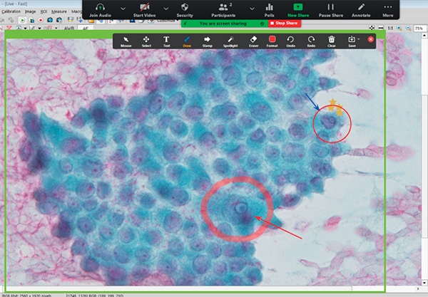
A variety of annotation tools are available on the Zoom platform (support.zoom.us/hc/en-us/articles/115005706806-Using-annotation-tools-on-a-shared-screen-or-whiteboard, accessed 12/05/2020). It is a good tool to engage the learners; questions can be asked that require annotating on the presenter’s screen (Fig. 1). Learners in cytopathology like the annotation resource because it allows for the teacher to point out morphological details precisely, better than using a pointer in a multiheaded microscope.
Another strategy that promotes active learning, made available by online sharing of the microscope images, is to “flip” sign-out, where the learner shares their screen and presents the case to the whole group. Recording the session can provide additional opportunities for self-reflection by the learner and individualized feedback by the teacher, remotely and at a convenient time.
While the pandemic has presented many challenges, the virtual format presents significant advantages, and the investment of time in learning to teach in virtual formats is a worthwhile pursuit. While we are all looking forward to getting back to in-person teaching and sign-out, we also hope to keep the best aspects of virtual learning to make educational opportunities more flexible and accessible for educators and learners after the pandemic is over.
- Mukhopadhyay S, Booth AL, Calkins SM, et al. Leveraging technology for remote learning in the era of COVID-19 and social distancing: tips and resources for pathology educators and trainees. Arch Pathol Lab Med. 2020;144(9):1027–1036.
- Kwon R, Zhang ML, VandenBussche CJ. Considerations for remote learning in pathology during COVID-19 social distancing. Cancer Cytopathol. 2020;128(9):642–647.
- Chiou PZ. Learning cytology in times of pandemic: an educational institutional experience with remote teaching. J Am Soc Cytopathol. 2020;9(6):579–585.
- Stathonikos N, van Varsseveld NC, Vink A, et al. Digital pathology in the time of corona. J Clin Pathol. 2020;73(11):706–712.
- Khan A, Egbue O, Palkie B, Madden J. Active learning: engaging students to maximize learning in an online course. Electronic Journal of e-Learning. 2017;15(2):107–115.
Dr. Kraft, a member of the CAP Cytopathology Committee, is associate professor and cytopathology fellowship program director, Virginia Commonwealth University, Richmond. Dr. Sundling, a member of the CAP Cytopathology Committee, is associate director of the cytology section at the Wisconsin State Laboratory of Hygiene, faculty director of the University of Wisconsin Cytotechnology Program, and assistant professor at the University of Wisconsin School of Medicine and Public Health.