


Anne Paxton
January 2025—Pathology informatics leaders at the University of Michigan are moving in steps to a fully digital practice as they put in place an innovative workflow for primary diagnosis.
Fresh off their August launch of the new workflow program within their remodeled histology lab, informatics directors in the pathology department say Michigan Medicine is the first in the U.S. to have radiology and pathology operating in the same shared picture archiving and communication system, or PACS, and to implement the well-established DICOM standard for pathology workflow.
“The unknowns that legitimately existed maybe five or 10 years ago”—whether storage and computation are adequate and whether the images are of adequate quality—“those have been answered,” says Ulysses G. J. Balis, MD, associate chief medical information officer and director of the Michigan Medicine Division of Pathology Informatics.
The expense will always need to be addressed, he admits. “This will cost money. That’s probably the biggest barrier right now. But the technology or the knowledge that it works—those are not the barriers anymore.”
The pathology department’s transformation was intended to be part of the move to its current offsite facility six years ago, though at the time they didn’t know when it would happen. “We knew at some point this building would be going to pure digital workflow,” and all the anatomic pathology spaces were designed for it, says Dr. Balis, who is the A. James French professor of pathology informatics and director of the computational pathology lab section.
He explains the physical reconfiguration of Michigan’s sign-out rooms. “The microscope used to literally be front and center in our sign-out spaces. The pathologist was seated centrally, with learners huddled nearby at adjacent microscope binocular heads. For this new workflow, we wanted to keep the microscope convenient but off to the side, so we had to change the configuration of our multiheaded scopes. While the microscope is still at the front and within arm’s reach, what is now front and center is the diagnostic-grade monitor, where most diagnoses are now made.” In addition, they have subordinate monitors throughout the room so fellows and residents can also see the cases conveniently.
“So we now have multi-monitor-equipped rooms, with the microscopes still multiheaded, but the primary manner in which cases are expected to be signed out is via use of a glass cockpit,” as he puts it, “with its associated diagnostic-grade computer monitor.”
At many institutions, space is at a premium and sign-out can be a “cramped and claustrophobic setting,” Dr. Balis says. “By virtue of being a new facility, when we designed these spaces in 2017, we knew we would be eventually transitioning to primary diagnosis by digital workflow and therefore purposely allocated far more space for sign-out rooms than was the norm of the day. In realizing digital workflow, having this space available has indeed paid substantial dividends.”
With the department’s transformation to a digital primary diagnosis workflow will come simplified interdepartmental case sharing and rapid deployment of advanced artificial intelligence solutions, he says. The next major phase of the project, to take place in the next 18 months, is to activate all of the subspecialty AP services in phased steps.
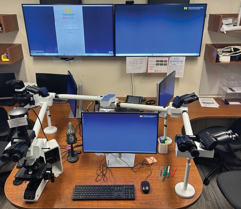
For the six-month transitional period during which slides will be co-delivered while whole slide image use for primary diagnosis is ramped up, a multiheaded microscope will remain available. However, a fundamental configurational change is evident in that the primary microscope viewing location is now sitting to the side location of the attending pathologist’s place setting, with the primary workplace now reserved for a medical-grade display. Additional display screens are available for the AP lab information system, gross images, electron microscopy images, electronic health record access (Epic), and the endoscopy image management system.
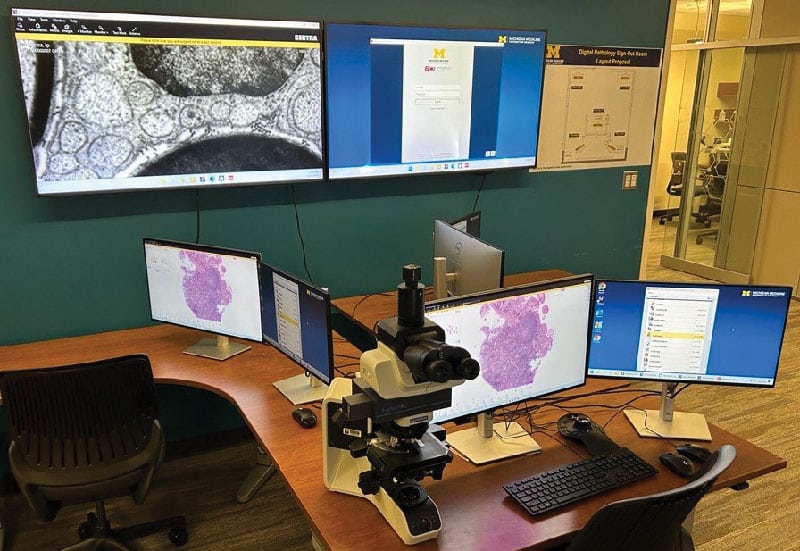
In the anticipated final production sign-out room layout, only a single-headed microscope will remain in place for the attending pathologist’s occasional use. The central monitor is now replaced by two centrally placed diagnostic-grade monitors, and further duplicated on the left and right with paired monitors that have exactly mirrored views, in support of resident and fellow teaching. A 3Dconnexion SpaceMouse is used for whole slide image navigation.
Michigan’s pathology department had an edge because 20 years ago, led by Bruce Friedman, MD, emeritus professor at Michigan, the informatics department’s pursuit of software development helped the department self-determine its future capabilities to conduct high-tech ventures involving software development and information technology within pathology itself, says Dr. Balis, who joined the team in 2006.
“This is uncommon among pathology departments, which often witness their IT being subsumed among the enterprise’s central IT group.”
“Michigan’s secret sauce, if you will, is that we’ve retained a substantial critical mass of local IT capabilities, not just for support but also for research and development,” he explains. “So we build our own software, we do original application development, and we do field-testing of candidate applications with our various user communities.”
The creation of a digital primary diagnosis workflow is a multimillion dollar investment for a department of Michigan Medicine’s volume, Dr. Balis says, and justifying it is the potential of AI and machine learning to have a positive impact on the level of care provided to patients.
“Now that the field of pathology is able to gather spatially anchored transcriptomics data—all the molecular data that can now be spatially aligned, if you will, with histology—this will allow for far more precise diagnosis and prediction of the biologic potential for a given individual. So you will be able to look at an image taken in tandem with that exact tissue’s associated pattern of molecular data, put it through one or more computational pipelines, and arrive upon a predictive set of outcomes for a patient’s biological potential for, let’s say, a tumor, to ask what will that tumor do at six months, a year, two years, in terms of its metastatic potential or recurrence potential. And if you compare and contrast that predictive capability with what you can currently do with the use of a microscope alone, there’s no comparison.”
In the U.S., he says, “We’re right on the cusp of the pathology specialty as a whole awakening to the fact that digital workflow for primary diagnosis is transitioning from a nice-to-have to a must-have. It should no longer be viewed as a luxury or a curiosity.” Pathologists should share with all colleagues what works and what doesn’t, he adds, “so people don’t repeat mistakes.”
Mustafa Yousif, MD, a breast and gynecologic pathologist at Michigan and director of digital pathology, points to the importance of the DICOM format for compatibility and integration with other departments, and for flexibility in equipment, the ability to upgrade without compatibility problems, and avoiding the inefficiencies of proprietary formats.
Michigan had a dozen years ago what many pathology departments have now—a standalone IT system, especially for digital pathology, to use for multiple purposes, including research, archiving, education, and consultation, Dr. Yousif says.
The weakness was that the IT has never been available for full clinical diagnosis. “The entire system was only connected to the Department of Pathology. We could not share our images with our colleagues or do multidisciplinary collaboration between radiologists, pathologists, and oncologists to review a case.”
DICOM adoption has helped make Michigan’s previously standalone digital pathology system part of the clinical enterprise imaging solution, where it’s integrated with radiology, Ob-gyn, and cardiology. Now, “We are part of any service or department that uses the medical image for diagnosis. And technically that means using DICOM format for diagnosis,” Dr. Yousif says.
The radiology department’s experience was an important factor in the pathology department’s adoption of its digital diagnosis workflow. What made the project successful was that pathology didn’t implement de novo the PACS product chosen (Sectra, made by Swedish firm Sectra AB), Dr. Balis says. Sectra, which is a contraction of “secure transmission,” “already was the enterprise’s vendor-neutral archive for images across the entire Michigan Medicine health system. We were just adding on a relatively small incremental module, which is pathology, and they had very good features for pathology. The storage, the PACS communication, the cloud-based aspects—all of that was already worked out by radiology.” That made it possible for pathology to focus on using the image management platform “to get cases signed out efficiently and quickly, with all the information you need locally available,” Dr. Balis says.
Eventually, the department’s storage of digital pathology data will be entirely in the cloud. “We’re not pure cloud yet,” he notes. “We have a lot of on-premises solutions still, because the size of a typical whole-slide image is large and therefore the computer needs to be near the stored data to allow for rapid image retrieval times. Therefore, for the time being, our primary storage is on premises.”
Use of a picture archiving and communication system-driven workflow, instead of the laboratory information system-driven workflow, is one of the keys to pathology’s new workflow. “This is the first time we are partnering with radiology and others to utilize the PACS,” Dr. Yousif says.
A longstanding obstacle to this kind of integration has been the proprietary nature of digital pathology components, he notes. “Making the contract with one vendor, you are stuck. You are locked with that vendor,” which will provide the scanner, server, and image viewer, “which can only work with one vendor.”
“If you want to do any collaboration”—for example, if one vendor provides only bright-field scanning but not immunofluorescent or polarization scanning—“each vendor has its own proprietary format. To go fully digital, you need to digitize all those aspects; you cannot just scan,” Dr. Yousif says.
“This is the setback. This is the problem we are living with on a daily basis,” he adds. “Right now we scan all these standard, one-by-three-inch slides. But you need a specialized scanner to scan those really large slides, and that means another vendor with a different proprietary format. On top of that, we have frozen section slides that are difficult to scan from the beginning. If one vendor you are working with does not have the capability of manual scanning, to add multiple focal areas for the scanner to start scanning in different planes, then your scanner is stuck. You are always going to have a blurry image from frozen sections.”
The problem is the lack of a standard format, Dr. Yousif says. Instead, the laboratory may use TIFF, BIFF, Mirax, SVS, or another format. “There are maybe 15 of these formats at the moment,” he says. In theory, if all of them used DICOM, that would solve the compatibility issue, and that is how it has worked at Michigan Medicine. “Right now, we have around 14 scanners from four different vendors, but all of them are now using the DICOM image format. So this literally solved the entire problem.”
The PACS is capable of reviewing the DICOM image format. It is the international standard for medical images and all the related information that comes with them, and it’s been used in radiology for the past 20 years, Dr. Yousif says. “They solved the compatibility issue more than 20 years ago.”
With PACS, going forward, pathology will follow radiology’s lead by eliminating the need for multiple platforms. We want to “put everything together in one digital platform—or, as we call it, a digital cockpit—to view all digital images at one time, not have to minimize or use multiple windows or go back and forth.”
The infrastructure for the PACS platform “has already been built within the radiology system,” Dr. Yousif explains. “So I encourage institutions that are trying now to move into digital to start communicating with the radiology team. Ask them if they have a solution that can read DICOM pathology, specifically called slide microscopy modality for DICOM pathology.”
As one of the co-chairs of the DICOM Working Group 26, Dr. Yousif helps advocate for standardization through adoption of DICOM. “Every other year we do a lot of work to provide proof of concept that we can integrate DICOM with multiple scanners, we can integrate with multiple PACS or digital platform systems as well as multiple vendor-neutral archives, and we use it even right now for annotation or artificial intelligence applications.”
“Our problem is that nobody within the U.S. can share DICOM pathology for the clinical diagnosis. Right now we have the portal if they want to send some proprietary format.” However, that image by itself, called a naked image, has no patient information, no protected health information, and no medical record number, he explains. “There is nothing so that I can recognize the case. But with DICOM, the metadata can be embedded within that image.” If pathology departments were to use this pathway with DICOM, “it would at least reduce the amount of technology that every department has to put in their budget every year” to reinterpret incoming information, he says.
Within Michigan Medicine, the departments have deep knowledge of DICOM, Dr. Yousif says. “All we need is to push the scanner and the image viewer vendors to adopt standardization.” But machine learning and AI make this a critical moment, he cautions. “An institution might have only one AI-based application that can, for example, detect all the micrometastases or all the tumor invasion for a given organ system in a patient. But the specific input and output data formats in use by this proprietary application might not be generalizable, and this limitation is a substantial barrier to widespread adoption.” DICOM use will allow for greatly simplified deployment of such algorithms across all IMS platforms, he says, as the DICOM standard enforces algorithmic compatibility with its standardized image formats and structured data types.
The workflow differences between pathology and radiology are well known, Dr. Yousif notes, but multiple PACS are already available that can read pathology and radiology. “One is Sectra, which has already been validated, and the FDA has also cleared another scanner for DICOM pathology for clinical use.”
“The rollout of the pathology department’s new workflow was one of the cleanest and most uneventful go-lives I can remember,” Dr. Balis says. Dr. Yousif, who led the rollout, gives some credit to the multiphase process they are using into January and beyond: “We are scanning 100 percent of our slides, every surgical pathology image, from bright-field, from immunofluorescence, from whole mount, from frozen section, polarization scanning, and also they have a PACS which is immunohistochemistry staining. Everything is fully digitized.”
The physical logistics of converting to digital presented challenges.
The area to scan digital pathology slides was small, Dr. Yousif says. “Then we studied the entire infrastructure for scanning and decided to use a separate room and we built that room to be a little closer to the main lab. Then someone has to send us those glass slides and then we digitized. That was the plan more than 18 months ago.” However, they then decided to visit multiple labs that already moved to a digital workflow. “And we asked, ‘What is the single thing that if you wanted to redo this, you would do for your lab?’ And they said, ‘I wish I could put my digital pathology scanner within histopathology or within the wet lab.’”
So Michigan Medicine decided to switch from the histology lab infrastructure installed in 2018 in an offsite location. “We said we want to remodel to have the digital pathology lab with 13 to 15 scanners in the center of the lab. So we relocated these scanners to make it efficient. The slides are distributed from staining direct to the scanner, then sent to the back counter for distribution without interruption, without interference or multiple floors or handling. And that’s helped us have extra space for different microtomy specimen devices” and streamline the entire workflow.
Hiring multiple FTEs to do extensive quality control is a necessity, Dr. Yousif says. “At this moment we are performing 100 percent QC for every slide. Every slide will have multiple stages of QC: a QC stage on the scanner, then a QC stage on the PACS, and then a QC stage between the block and the glass slide, just to make sure the entire tissue being captured, the entire area, is not out of focus.”
All this QC means manual labor and added costs, but Dr. Yousif predicts that QC artificial intelligence will soon help, made possible by DICOM. “With an AI application, it can tell you even before you take the slide from the scanner that slide number two in the rack or number five in the scanner has issues and you need to re-scan that slide.” This isn’t feasible with a proprietary format other than DICOM, Dr. Yousif says. “This is available because DICOM provides you all this metadata or granularity of detail to find that slide.”
The histotechnologists love this feature, he adds. “You have to provide tools or data to help your histotechs. We created dashboards for them because the visualization is important for the scanning histotech as well as for the other histotechs.” This way, they can say, “‘Oh, I see from the dashboard that scanner number one is done with staining so I need to pick it up.’ It tremendously improved workflow to have the dashboards provide this notification.”
He advises anyone with doubts about advancing their digital pathology to take a look at sites where it is working. “Visit multiple sites that have success stories.
Communicate with them; learn from their mistakes. Then the first thing is to try to communicate with your colleagues from radiology, from their imaging department, because they might have a solution for you.”
When he and his colleagues presented their plan to radiology, “they had no idea about pathology. We educated them and they helped us.” Now “we are part of their system. So when we hire additional FTEs, they are helping radiology, and their FTEs are helping us.”
Second, he advises putting DICOM into the contract with any vendor so the vendor is responsible for paying for all the conversion, all the migration. “Then the vendor is committed from now on.”
His third piece of advice is to use what is called a unified platform. “For example, think about what’s called X-driven workflow or a platform or digital cockpit that can help pathology.”
Finally, Dr. Yousif says, an important question is how to make your digital pathology information available to the patient and clinician. “We are living in this bubble by ourselves and sharing all this knowledge for patient care. So if my patients right now wanted to transfer the care from Michigan Medicine to another institution, different state, or even a different country, how can I make it efficient by sending the data so the receiver can read it? Again, with DICOM, because everyone understands the standard, they don’t have to install another application or work with that vendor to decipher that format. You send the data in an encrypted way to the other institution and they should be capable, to be honest, of reviewing those images.”
His hope is that at the end of 2025 the department will be fully digital for all surgical pathology services. “By that time I’m hoping we barely distribute any glass slides because [for now] we have this hybrid workflow.” The different services are pleading with him not to send any more glass slides, he says. “Pediatric, renal, medical—that’s literally what they have said.”
Nevertheless, building a digital pathology workflow also has to take into account the capability to pull slides, Dr. Yousif says. “In the PACS-driven system, we have built all the customization that can enable the pathologist to request glass slides if they need to.”
The new workflow includes a backup plan for disaster recovery, ensuring continuity in the event of system downtime. Another PACS allows images to be saved for just seven days. After that, “we purge it from the backup PACS because you don’t need it because the main one is already archived.”
At some institutions, he says, the likely backup plan is to distribute glass slides during downtime. “And that’s fair enough, but we need to change this mindset. I want to provide everything I can in order not to go back to the glass slide.”
Dr. Yousif asks colleagues at other institutions if they’re able to provide their images to their clinicians. “There is a lot of fear about doing that, but sharing our knowledge and our images with clinicians and with patients directly is a patient care and patient satisfaction-enhancing opportunity,” Dr. Yousif says. “The interaction allows the clinical team to more precisely tailor treatment regimens and, similarly, allows the patient to better understand their own disease process.”
From the experiences gained from piloting trials at Michigan, Dr. Balis says, “we know it is highly impactful and beneficial when patients get the opportunity to review their histology with a pathologist. It’s not standard practice at all yet. It’s something we aspire to do. But from the limited examples we have, we know it works.”
Anne Paxton is a writer and attorney in Seattle.