

Amy Carpenter
May 2025—How the World Health Organization fifth edition of hematolymphoid tumors and the International Consensus Classification differ for myeloid malignancies was highlighted in cases presented in a CAP24 session last fall.
Second of two parts
Part one in the April issue
Sanam Loghavi, MD, associate professor of pathology and laboratory medicine, Department of Hematopathology, University of Texas MD Anderson Cancer Center, spoke of myelodysplastic neoplasms/syndromes (MDS) with defining genetic abnormalities and the allelic state of TP53 in MDS, among other things. (Kamran M. Mirza, MD, PhD, of the University of Michigan, co-presented. See part one in the April issue: https://bit.ly/CT_0425-myeloid.)
Dr. Loghavi’s first case was that of a 51-year-old woman who underwent a workup for pancytopenia. The patient’s CBC showed absolute neutropenia with a low white blood cell count (1.2 × 10⁹/L). She had anemia (Hgb: 9.8 g/dL) and thrombocytopenia (Plt: 41 × 109/L). Her MCV was normal (92 fL).
The bone marrow biopsy (Fig. 1) was fibrotic and hypercellular for age, Dr. Loghavi said. The reticulin stain (middle) confirmed the presence of fibrosis. Often in cases that have high degrees of fibrosis, “the aspirate smears are terrible. You cannot see dysplasia; it’s difficult to locate the blasts” without looking at many aspirate smears, which is what they did. Two blasts can be seen on the aspirate smear (right). “Overall there were 16 percent blasts but in a hemodiluted smear,” she said. (More on this case later.)
In the WHO fifth edition (WHO-HEM5) in general, “the genetic categories trump morphologic categories,” Dr. Loghavi said (Khoury JD, et al. Leukemia. 2022;36[7]:1703–1719).
One such genetic category is MDS with low blasts and isolated 5q deletion, which she calls “a misnomer because despite what the terminology implies, you can have one additional set of genetic abnormalities as long as it doesn’t involve chromosome 7,” because the latter is associated with a poor prognosis and del(5q) is typically thought to be a lower-risk disease. (In the WHO fifth edition, myelodysplastic syndromes were renamed myelodysplastic neoplasms—abbreviated MDS.)
Another genetically defined category of MDS in the WHO classification is MDS with low blasts and SF3B1 mutation (MDS-SF3B1). The variant allelic frequency qualifier is one difference between the MDS with SF3B1 mutation categories in the WHO and ICC classifications (five percent, WHO; 10 percent, ICC).
The WHO allows for a branching category of MDS with SF3B1 mutation, Dr. Loghavi said. “If you don’t have a mutation or are in a resource-poor setting without [access to] sequencing and you have more than 15 percent ring sideroblasts, you can still put the case in this diagnostic category, but instead of calling it ‘with SF3B1 mutation,’ you call it MDS with ring sideroblasts.” The reason: In the initial trials studying response to luspatercept [Reblozyl], patients with ring sideroblasts and SF3B1 mutations benefited the most from the drug. “For the ICC,” Dr. Loghavi said, “you need to have the mutation and only that is recognized as a distinct category.”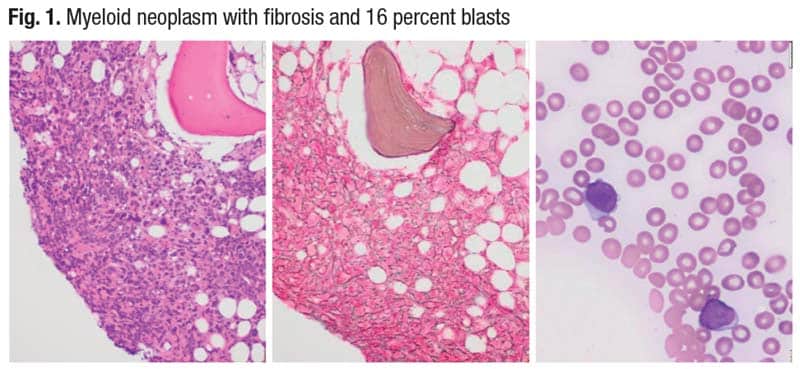
The third category of MDS with defining genetic abnormalities is MDS with biallelic TP53 inactivation (for now, it is not AML-defining in WHO). Its defining features in WHO are less than 20 percent blasts in bone marrow and peripheral blood, usually complex cytogenetics, and two or more TP53 mutations or one mutation with evidence of TP53 copy number loss or copy neutral loss of heterozygosity. The WHO uses the term biallelic, and the ICC uses multi-hit, “which essentially means you lose both functional copies of TP53 by different means,” Dr. Loghavi said. It’s her view that “the ICC approach is probably more accurate in this case” (Arber DA, et al. Blood. 2022;140[11]:1200–1228).
“If you don’t have any of these genetic categories, you’re left with a morphologic classification,” she said, and most important would be the blast count. “If you have more than five percent blasts or more than two percent in the peripheral blood, or more than 10 percent blasts and more than five percent in the peripheral blood, you’re in the increased blast category.” In the WHO classification, MDS with fibrosis is also a distinct morphologic category. If the bone marrow is severely hypocellular for age, it is hypoplastic MDS (≤ 25 percent cellularity, age adjusted).
The ICC does not recognize hypoplastic MDS. “Biologically there is overlap between hypoplastic MDS and aplastic anemia,” Dr. Loghavi said. They’re thought of as two different diseases and they are, “but there’s a biological spectrum,” and the evidence shows that some patients with hypoplastic MDS respond well to immunosuppressive therapies and not necessarily to hypomethylating agents. For therapeutic purposes, the WHO authors thought “they would pull this category out to give the patients a chance” for response on immunosuppressive therapies.
If the patient is not in any of these categories, and there is increased blasts, then it is MDS with increased blasts. Designation of morphologic dysplasia, whether unilineage or multilineage, is optional in the WHO classification.
The ICC is similar, she said. The major difference is that in the ICC classification the two morphologic categories—hypoplastic MDS and MDS with fibrosis—are not recognized as distinct categories.
One major difference in the way the ICC and WHO classify MDS and AML, Dr. Loghavi said, is that a 10 percent blast count in the ICC takes a case out of the classic MDS category and places it in MDS/AML. “So if you have between 10 and 20 percent blasts without an AML-defining genetic alteration, it’s MDS/AML.” The WHO says that for clinical trial enrollment and therapeutic purposes, MDS-IB2 (the equivalent of MDS/AML) can be treated as AML at the discretion of the treating physician if the physician thinks the patient will respond better to AML-type therapy.
Why does the allelic state of TP53 in MDS matter?
A fortuitous finding came about as part of the International Prognostic Scoring System risk stratification project, Dr. Loghavi said (Bernard E, et al. Nat Med. 2020;26[10]:1549–1556). In patients who had multi-hit TP53 mutations, leukemia-free survival and overall survival outcomes were particularly dismal. Bernard, et al., write, “Surprisingly, monoallelic TP53 patients did not differ from TP53 wild-type patients with regard to overall survival and AML progression.”
Given the biology of TP53, “this makes perfect sense because TP53 is a tumor suppressor gene,” Dr. Loghavi said. “Typically in any type of malignancy, when there is a dysfunction of a tumor suppressor gene and you have one conserved copy, you normally don’t have a catastrophic event because the normal copy compensates for the abnormal copy. But once you lose both copies of a tumor suppressor gene, you lose the function of that gene. That is what happens with TP53.”
There are three ways to get to biallelic or multi-hit status, Dr. Loghavi said, first explaining her view that the multi-hit terminology is better. “Although on the science side we’re very sophisticated, in clinical practice nobody is doing single-cell sequencing. We’re doing bulk NGS, so there’s no way for us to know this is in fact a biallelic hit even if you have two mutations, unless the mutations are so close to one another physically that we can see them in the same reads in the NGS results. But we’re inferring that if there are two mutations, they’re in two different alleles because we know the biology of TP53.”
If there is one mutation and the other copy is lost by way of deletion, it can be seen on a routine karyotype or by FISH for TP53. MD Anderson Cancer Center, probably like most academic practices, she said, does not routinely perform FISH for TP53 unless “we see it on a karyotype, if we see an abnormality on 17p.” If there is a TP53 mutation at a high VAF or a complex karyotype with a TP53 mutation, “we will add a TP53 FISH to make sure we’re not missing a TP53 deletion.”
The most challenging scenario is having a copy neutral loss of heterozygosity of TP53 with a mutation, she said. The mutation is seen on the sequencing. But in most clinical laboratories, the assays don’t have the ability to detect copy neutral loss of heterozygosity. “This is the same kind of mechanism as a uniparental disomy. You have one mutant copy, and then the normal copy is deleted but the mutant copy is duplicated. So if you FISH for TP53 you’re going to see a normal signal because you have two copies of TP53 but both are abnormal.”
“If you have one mutation with a variant allele frequency of more than 50 percent,” she continued, “you can assume that the wild-type copy is deleted. You’re inferring and using it as a surrogate, but it normally works pretty well” (El Hussein S, et al. Cancers (Basel). 2022;14[22]:5690).
At MD Anderson, pathologists run a p53 immunohistochemistry on every newly diagnosed case of MDS or AML. “It’s much faster than sequencing and it’s a good screening tool,” Dr. Loghavi explained. A normal pattern of p53 staining is typically faint and patchy nuclear staining (Fig. 2, top left). “When you have a mutation—typically a missense or a splice-site mutation—the mutant copy is resistant to MDM2 degradation, so you get accumulation of p53 in the cell and the antibody picks up much higher levels of p53, and there is intense, dark nuclear staining [top right]” (Tashakori M, et al. Blood. 2022;140[1]:58–72).
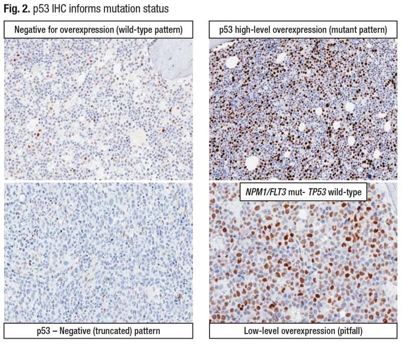
Staining intensity is important. “Moderate is not what we’re looking for,” Dr. Loghavi said. “It’s very bright, intense staining. And the number of cells that stain typically is commensurate with the variant allelic frequency of the mutation.” The higher the VAF, the more cells that stain intensely.
Another abnormal pattern—the null pattern—typically goes with a nonsense or early terminating frameshift mutation that results in the antibody not recognizing the protein (bottom left). There is weak staining in the spindle cells which are the endothelial cells, but the myeloid cells are negative.
A p53 IHC stain is agnostic of the allelic status and thus won’t help in the classification of MDS. “You can say whether there’s a mutation or not, but you can’t tell if it’s biallelic or multi-hit,” she said.
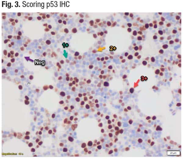
In scoring a p53 IHC (Fig. 3), “the only intensity we care about is 3+ [red arrow],” Dr. Loghavi said. If more than 10 percent of cells are bright, intensely staining cells, there likely will be a mutation at a VAF of 10 percent or higher.
She returned to the case of the 51-year-old patient with increased blasts and fibrosis. Most important is whether there is a TP53 mutation, and there was a TP53 mutation at a variant allele frequency of 60 percent, she said, and a complex karyotype including del(17p). In the WHO classification, the case is MDS with biallelic TP53 inactivation (biallelic by definition because of the mutation and the 17p deletion). “Even though you have fibrosis, TP53 status trumps the presence of fibrosis,” Dr. Loghavi said.
In the ICC, because there is 16 percent blasts—more than 10 percent—the case would qualify as MDS/AML with mutated TP53. Patients with more than five percent blasts have a poor prognosis and should be considered for allogeneic stem cell transplant if possible, she said.
The WHO classification does not recognize myeloid neoplasms with TP53 mutation as a distinct category. “Most AML cases are classified as AML with myelodysplasia-related cytogenetic abnormalities by virtue of the complex karyotype they are typically associated with,” Dr. Loghavi said. ICC recognizes MDS/AML with mutated TP53, “and you don’t need a biallelic or multi-hit status. Any TP53 mutation with a VAF of more than 10 percent will qualify for this category.” AML with mutated TP53 is identified by more than 20 percent blasts and any TP53 mutation with a VAF of more than 10 percent.
“These patients are going to have a very short window between when you can get them into remission and into transplant,” she said.
She cited one of many publications that showed “when you have a myeloid neoplasm with a TP53 mutation with an increase in blasts—more than five, 10 percent—regardless of the allelic status and the blast count, the patients are going to do equally as poorly” (Grob T, et al. Blood. 2022;139[15]:2347–2354).
Dr. Loghavi presented a second case, that of a 71-year-old man who underwent a workup for pancytopenia (Loghavi S, et al. Mod Pathol. 2024;37[2]:100397).
The patient was neutropenic (WBC: 1.5 × 10⁹/L) and had a low leukocyte count and low hemoglobin (8.9 g/dL). He was severely thrombocytopenic (Plt: 6 × 10⁹/L). His MCV was 96 fL.
In the core biopsy, distinct, large cells with crisp, nuclear outlines and carrot-like nucleoli stuck to the membrane were a typical picture of erythroid blasts, Dr. Loghavi said. “All of the hematopoiesis is replaced with erythroid blasts, confirmed on the aspirate smear,” which showed intensely blue cells with round, punched-out nuclei and vacuolated cytoplasm. The CD71 stain confirmed erythroid differentiation. The p53 stain revealed an abnormal mutant pattern. “We had 82 percent erythroid cells, 42 percent of which were erythroid blasts.” (More on this case later.)
The majority of AML cases are classified by their genetic alteration, both in WHO and ICC. “When I’m on an acute leukemia service, if I see 10 AMLs, I’m able to genetically classify nine of them,” Dr. Loghavi said. “It’s rare to come across a case that’s not genetically classified and [have to] go by morphology.” But those rare cases exist, she added, and she suspects the genetic alteration is not being detected or is nondescript. In WHO-HEM5, several alterations are AML-defining, among them the NPM1 mutation.
What’s important, she said, is that the 20 percent minimum blast count requirement for AML has been removed in the WHO classification. An increase in blasts is required, “which is essentially five percent or higher.” The case qualifies as AML with one of several genetic abnormalities listed.
But the 20 percent blast count requirement remains for AML with CEBPA mutation (in WHO) or with BCR::ABL1 (in WHO and ICC). “You don’t want to overdiagnose CML in accelerated or blast phase as a de novo AML, so AML with BCR::ABL1 is restricted to de novo AML cases,” Dr. Loghavi said. “And for CEBPA, the WHO thought there was not sufficient evidence to say that cases with less than 20 percent blasts behave similarly to those with a higher blast count. But in routine practice when you have a pathogenic CEBPA mutation, the blast count is typically high, so that dilemma is not much of a dilemma.” The 20 percent requirement also remains for myelodysplasia-related AML (AML-MR).
If there is no defining genetic abnormality, differentiation is used.
It’s Dr. Loghavi’s view that it was a mistake to completely replace phenotype with genotype in the newer classifications. “Phenotype still speaks to the biology of disease,” she said, “and there’s evidence to support that.” Monocytic, erythroid, and megakaryocytic leukemias don’t respond as well to venetoclax, in part because they’re enriched in TP53 or RAS mutations that make these leukemias resistant to BCL-2 inhibitors. “The differentiation is important, and we should put that in our top diagnostic line. You can have an NPM1-mutated AML and call it that, but if it’s purely monoblastic you want to indicate that and say, ‘This is an NPM1-mutated AML with monoblastic differentiation.’”
The ICC is similar, she said, with the difference being that the AML genetic qualifiers still require 10 percent blasts, including for acute promyelocytic leukemia and core-binding factor AMLs. “In routine practice, it’s not much of a dilemma if you have an APL or a core-binding factor because the blast count is usually more than 10 percent,” Dr. Loghavi said, but a few cases may not meet the 10 percent blast count threshold. In addition, the ICC doesn’t use differentiation. “They call everything AML NOS.”
Back to the case of the 71-year-old patient with pure erythroid leukemia. In the 2016 WHO classification, there was such a category, which is an extreme erythroid differentiation in the blasts. Many publications support the association of pure erythroid leukemia with multi-hit or biallelic TP53 alterations, Dr. Loghavi said. This leukemia is usually seen with a TP53 mutation, a complex karyotype, and a high VAF. “It’s essentially defined by the presence of TP53 mutations,” she said.
The 71-year-old patient had extensive erythroid differentiation and a TP53 mutation. “In the WHO fifth category, even though this has TP53 mutation, the presence of erythroid differentiation supersedes AML with myelodysplasia-related changes,” Dr. Loghavi said. “You would call this an acute erythroid leukemia.”
In the ICC, by virtue of the presence of the TP53 mutation and alterations, all these cases are lumped into AML with mutated TP53. “Even if you want to call it AML with mutated TP53, it’s helpful to put the erythroid differentiation in the diagnostic top line,” she said.
Are TP53 mutations universally bad? The context in which they occur matters, Dr. Loghavi said (Stengel A, et al. Blood Adv. 2023;7[13]:2952–2956). In the majority of TP53-mutated cases, mutation frequency confers a bad prognosis. But when the frequency of TP53 mutations is not high or when the TP53 mutation co-occurs with a mutation like an NPM1 mutation, or in a core-binding factor or an APL, “it usually doesn’t have the same awful prognosis that the other cases do,” though the number of cases is low so the studies are not extensive, she said. When such a case is seen, she advises conveying the difficulty of the classification, noting the presence of co-occurring good and bad mutations, and explaining that the significance of the prognosis is unknown.
In the third case Dr. Loghavi presented, a 61-year-old female with a past medical history of breast cancer treated with chemotherapy undergoes a workup for cytopenia that identified a TP53 mutation (p. D281G) with a VAF of 76 percent.
(The TP53 mutation is multi-hit because the 76 VAF is more than 50 percent, she noted, adding, “Even if there’s no deletion, this has copy neutral loss of heterozygosity.”)
The CBC reveals pancytopenia: WBC: 2.3 × 10⁹/L absolute neutropenia, Hgb: 6.9 g/dL, Plt: 25 × 109/L, and MCV: 90 fL.
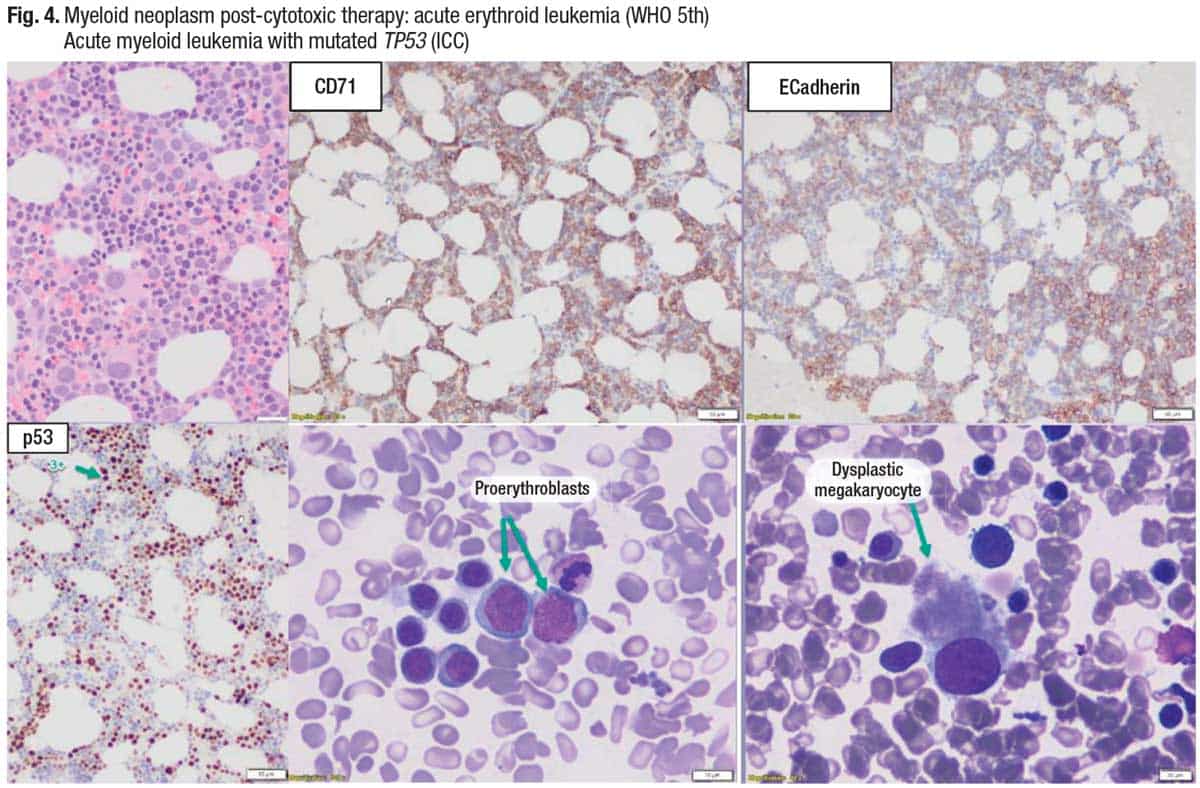
The bone marrow biopsy (Fig. 4, top left) shows that the megakaryocytes are dysplastic, and there is erythroid differentiation (CD71). Immature erythroid cells are highlighted (E-cadherin), “so you can see these are erythroid blasts.” P53 shows an abnormal pattern of staining, and the two aspirate smear images (bottom middle and right) show increased erythroblasts and a dysplastic megakaryocyte.
“One of the subtle differences in the classification of myeloid neoplasms in the ICC and WHO is that in the WHO, therapy-relatedness trumps the classification,” Dr. Loghavi said. “In the ICC, classification trumps therapy-relatedness.” She prefers the ICC approach “because even if you have a patient who has been exposed to chemotherapy, if they have an AML with NPM1 mutation or an APL, that’s probably not going to behave like a therapy-related myeloid neoplasm.” But most cases are post-therapy and have bad genetics, such as a TP53 mutation or KMT2A rearrangement, and will behave poorly, Dr. Loghavi said. In her practice, she first qualifies the case genetically and then will say the patient has a history of exposure to cytotoxic therapy. “That does both classifications justice and leaves the physician with treatment options.”
In the case of the 61-year-old female patient, there is erythroid differentiation and thus the case would be classified as acute erythroid leukemia in the WHO classification.
On the issue of therapy-relatedness, Dr. Loghavi cited the clonal hematopoiesis risk score. It provides a prognostic framework for clonal hematopoiesis of indeterminate potential and clonal cytopenia of undetermined significance by distinguishing a high-risk minority from the majority of CHIP/CCUS, which has a minimal risk of progression to myeloid neoplasm (Weeks LD, et al. NEJM Evid. Published online April 25, 2023. doi:10.1056/EVIDoa2200310).
“If healthy individuals who have a TP53 mutation are not exposed to chemotherapeutic agents, you don’t disrupt the environment, they can live a long time with a high-VAF TP53. It will just sit there,” Dr. Loghavi said. “For reasons that are biologically unclear to me, when you do the stain, you know there is a mutation, but it seems that the cells are able to discard the mutant p53, so you don’t see an abnormal pattern of staining. But once you expose these patients to chemotherapy, then you select for these TP53-mutated clones because you’re killing the wild-type cells, but these cells don’t undergo apoptosis. So you’re selecting for them and waiting for the second hit to happen. Once the other copy is deleted or mutated, it explodes. This is how you get therapy-related myeloid neoplasms enriched for TP53 mutations compared with de novo TP53-mutated neoplasms.”
Does the allelic status matter in the setting of TP53-mutated myeloid neoplasms? “At least in one study, if you have a therapy-related myeloid neoplasm, it doesn’t matter if you’re multi-hit or single hit,” Dr. Loghavi said. “You’re going to do poorly. The only thing that matters is the burden of the clone” (Hiwase D, et al. Blood. 2023;141[9]:1087–1091).
In the case of a therapy-related myeloid neoplasm with a TP53 mutation, as long as the VAF is less than 10 percent (in practice such cases are rare, she said), patients may not do as poorly, Dr. Loghavi said. “You can postulate that maybe the therapy-related neoplasm, in the presence of a VAF less than 10 percent, is not really the driver of the disease. Maybe the driver of the disease is something else.”
Amy Carpenter is CAP TODAY senior editor.