
Karen Titus
March 2025—Like an inspired Adam in the Garden of Eden, molecular experts have been busy with the naming process as it applies to liquid biopsy.
It’s lost to the myths of time whether Adam revised his nomenclature, but pathologists and other experts are eager to identify new assays as they push this field forward, from circulating cell-free DNA to circulating tumor DNA to circulating tumor RNA.
Soon another assay, one that combines ctDNA and ctRNA, could begin to make its mark as well, says Keyur P. Patel, MD, PhD, medical director of the molecular diagnostics laboratory, Division of Pathology and Laboratory Medicine, University of Texas MD Anderson Cancer Center. He and his colleagues plan to launch an assay to look for circulating total nucleic acid. “DNA plus RNA equals TNA—that’s the mathematical equation,” jokes Dr. Patel, who is also professor, Department of Hematopathology, Division of Pathology and Laboratory Medicine.
And like that first zookeeper, there’s even an actual beast for experts to name. “Of course, the biggest elephant in the room is the reimbursement for all this,” says Shashikant Kulkarni, PhD, deputy division head and professor of molecular pathology and cytogenetics, Division of Pathology and Laboratory Medicine, MD Anderson. The Centers for Medicare and Medicaid Services pays for some liquid biopsy tests for some advanced cancer tumor types, but not for all, and private insurance coverage varies significantly, he says. “It’s very complex. And the landscape is evolving every day.”
The same can be said of liquid biopsy’s potential uses. Marching alongside these developments are emerging applications, as researchers look to extend early studies to clinical practice. Studies have already shown its ability to classify patients into risk categories and select treatment. Monitoring disease progression and treatment response also appears promising, “though we still need more information to match it with actionability of these emerging clones,” Dr. Kulkarni says.
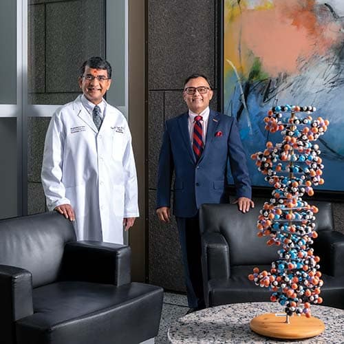
Dr. Shashikant Kulkarni at MD Anderson Cancer Center. “Clinical utility and interpretation are not straightforward,” Dr. Kulkarni says of the liquid biopsy. “We need more trials. We need more controlled studies; more longitudinal
evidence gathering is needed.” [Photo: Hall Puckett]
Even with so many unknowns, liquid biopsy has become a go-to test in many situations, says Dr. Kulkarni. At MD Anderson, it’s valuable when the patient’s primary diagnostic specimen is exhausted or not available and the oncologist requires further follow-up. “And there are scenarios where the patient has a metastatic tumor and it’s not clear what tissue should be biopsied.”
It’s also valuable because with its faster turnaround times, it can enable earlier treatment.
Liquid biopsy is crucial, in fact, and studies have shown strong concordance between tissue testing and liquid biopsy. “Of course, there are a lot of caveats to that,” Dr. Kulkarni adds.
That said, tissue remains the gold standard, and liquid biopsy is used as an adjunct when tissue is not available or there is a rapid need. “Even if they find an actionable marker,” Dr. Kulkarni says, “the oncologist usually prefers to get a tissue baseline readout as well.”
The term liquid biopsy has different meanings for different people, though at its essence, it refers to looking at the material from the traditional tumor tissue in peripheral blood.
Other samples are being looked at as well, says Dr. Kulkarni, including urine-based liquid biopsy, pleural fluid or ascites, and saliva. Researchers, including those at MD Anderson, are also exploring the use of cerebrospinal fluid.
Circulating DNA offered the first toehold, and it remains the primary approach for many laboratories that offer this testing. Studies on DNA outpace those on RNA, in part because DNA is considered to be the more stable of the two, Dr. Patel says. “So we know more about DNA than RNA in circulation, and that has become the more common target for different liquid biopsy applications.”
Changes at the DNA level include point mutations or small insertions or deletions. Other primary changes at the DNA level include gene rearrangements, in which two different genes from different portions of the same chromosome or from different chromosomes altogether form a chimeric fusion that leads to oncogenesis.
These changes are carried into RNA, Dr. Patel explains. “So you can look for translocation at either level,” though change at the RNA level is sometimes easier to target. “You’re only looking for a specific junction of the RNA. Whereas at the DNA level, those breakpoints can be far apart.” Given that RNA-based approaches are more recent developments, liquid biopsy assays for the most part look for DNA-level fusions. But as the technology evolves, it should become easier to show RNA transcripts and portions of transcripts circulating in the blood.
Adds Dr. Kulkarni: “The mainstream approaches today are primarily looking at single nucleotide variants and fusions, in limited scenarios.”
While the fusions arise at the DNA level, the ability to detect DNA-level breakpoints is limited because the breakpoints can span a huge region of introns. It’s often not possible or practical to cover that much territory, so to speak. But when the chimeric DNA is transcribed into RNA, says Dr. Patel, matters become more simple. He uses the example of exon 6 and the EML4::ALK fusion gene, which is one of the fusions commonly seen in non-small cell lung cancer. The breakpoint is downstream of exon 6 of EML4 and upstream of exon 20 of ALK.
Once breakpoints occur, Dr. Patel says, the RNA transcription gets rid of intronic sequences, which are basically excess amount of sequence, “and you are left with two juxtaposed exons. So you know exactly where the breakpoints are: E6/A20.” That requires much less sequencing as part of the assay design. The DNA-level approach is not practical for all fusions, given labs do not currently have the resources for unlimited sequencing. But with RNA—depending on the technology that’s used—“you can catch many more fusions, using even less sequencing effort in theory.” Amplicon-based technology, for example, can target the two fusion exons with small probes.
Dr. Patel and colleagues at MD Anderson are trying to come up with an approach that entails total nucleic acid extraction—the aforementioned cTNA. “We split that into DNA and RNA. There’s very little material to work with—in the nanogram quantity—and we have to make the most of it. We look at DNA separately, and we look at RNA separately, but from the same common TNA extract. Then we can identify both pieces of information simultaneously.”
And in the not-too-distant future, says Dr. Kulkarni, methylation-based approaches may start to have an impact on the field, which could improve residual disease deduction accuracy.
Clinical applications are continuing to grow apace as the technology develops, particularly in advanced-stage tumors that can be difficult to biopsy by traditional means.
There are also FDA-approved indications forthcoming for additional tumor types, where liquid biopsy will be an acceptable investigation to identify a targetable mutation and to direct therapy. Says Dr. Patel, “It’s not universal across all tumor types, but depending on the evidence, for certain tumor types, such as thoracic non-small cell carcinoma, it is an acceptable application even for direct treatment selection.” One obvious advantage is that investigations can move more quickly than when scheduling an invasive biopsy procedure, thus speeding up treatment.
A negative result does not rule out the presence of an alteration, however, limiting the value of a liquid biopsy in certain situations. “If you don’t find any informative results, it cannot be concluded there are no informative findings,” Dr. Patel says.
In other words, the need for speed should be matched with careful forethought, Dr. Patel and others caution. Below the bright promise of liquid biopsy are biological basics that researchers are still sorting out.
The most important specimen issue to bear in mind, Dr. Kulkarni says, is that each tumor type differs in the extent to which they shed nucleic acid into the bloodstream.
“That’s a pretty significant variable,” he says. High-shedding tumors include non-small cell lung cancer and colorectal, pancreatic, breast, and esophageal cancers. At the other end of the spectrum are brain tumors, where the blood-brain barrier limits the amount of ctDNA. Other cancer types, including sarcomas, thyroid tumors, and testicular and prostate tumors, are also low shedders. Shedding is primarily dependent on type of tumor, its size (large tumors shed more DNA), and vascularization. And metastatic tumors are more likely to release more ctDNA than localized tumors, as are more aggressive tumors.
“So even though we like to offer these tests in a pan-cancer way, we have to be very careful about false-negatives,” Dr. Kulkarni says. Conversely, clonal hematopoiesis of indeterminate potential can produce false-positives.
“There are things we are still learning at the individual tumor level,” says Dr. Patel. “We cannot generalize.”
He adds: “The majority of our observations are at one point in time. So we have highly variable correlations as the tumor burden increases.” If a patient has multiple different tumor metastases or loci, then the overall tumor burden is high, and the likelihood of some of these releasing DNA or nucleic acid material increases. “You tend to see that reflected in the actual liquid biopsy information in terms of the ctDNA fraction, in terms of the variant allele frequency of any mutation you identified.” That’s the reason why the majority of clinical applications home in on advanced metastatic tumors. “Essentially the liquid biopsy is an informative surrogate in those cases.”
In short, there are definite biological and clinical factors that will determine the assay’s utility, Dr. Patel says. “That’s why if we see a mutation in circulation, it’s always useful. But if we don’t see it, that doesn’t mean something’s not going on.”
Explaining test results, not surprisingly, comes with its own complications. At MD Anderson, the most commonly asked questions Dr. Kulkarni fields from his oncologist colleagues are about concordance—or lack of it—with tissue biopsy results.
Concordance between tissue and liquid biopsy also depends on the tumor type and stage of the cancer. Studies have shown concordance rates range from 60 to 90 percent. It’s lower when the tumor does not shed enough DNA, Dr. Kulkarni says.
Tumor heterogeneity can affect results, as well as timing of the sample. “Concordance is higher when you take the tissue and the biopsy at the same time,” he says. “Even a few weeks difference can affect the result,” because of tumor evolution and therapy-induced changes. Sensitivity of the analytical method—next-generation sequencing versus digital PCR versus droplet digital PCR—affects concordance as well.
Once the decision is made to use liquid biopsy—at least as a first step—preanalytical variables come into play.
“This is not an easy sample to work with,” says Mark Linder, PhD, professor of pathology and laboratory medicine, University of Louisville School of Medicine. “The concentrations are very low, and there has not been a lot of standardization on managing the plasma for extraction.”
That includes using the right collection tube. It may seem like an obvious point, especially as liquid biopsy has settled into clinical practice in recent years; moreover, testing is clustered primarily at academic medical centers with well-established resources and processes, as well as at commercial labs. “But it’s an important enough issue,” Dr. Patel says, “that anybody who’s just starting to do it should keep in mind that the type of collection tubes and preanalytical conditions will have bearing on the outcome.”

“Since you’re basically trying to find these few floating molecules in plasma,” he continues, “specimen stability becomes an issue.” If the blood cells aren’t properly stabilized, they may start to die and release their own DNA and other genetic material into the specimen, whereas a well-preserved specimen will contain only the tumor-derived DNA. The goal is to avoid the noise generated by nontumor-derived DNA. Otherwise, it becomes challenging to identify the actual signal, which is already low to begin with.
(Dr. Patel cites the “excellent” AMP/CAP joint consensus recommendation for cell-free DNA assay validation that labs can refer to: Lockwood CM, et al. J Mol Diagn. 2023;25[12]:876–897. “The experts painstakingly reviewed 1,228 circulating tumor DNA publications to develop a set of 13 best practice recommendations,” he says.)
If laboratories are unable to collect specimens in tubes that have stabilizing material, Dr. Patel says, then they will need to do their own studies and ensure that the specimens are ideally transported in conditions that allow as much plasma as possible to remain free from additional genomic material released from nontumor cells.
The specimens also need to be preserved in a timely manner, Dr. Patel continues. His group has published studies comparing use of EDTA tubes with those that use stabilizing material. For labs shipping specimens to an outside laboratory for testing, “That’s where sample stability becomes very important.”
“Preanalytical factors are still critical,” he adds. “Anybody who’s implementing this for the first time and putting together their workflow should be careful to make sure they’re implementing best practices.”
Those practices are far from settled. “This is a huge topic,” Dr. Kulkarni says, “and it’s ever-evolving. There is not enough standardization.”
Dr. Patel agrees, noting that the proprietary nature of much of the assay development has created one hurdle. “We need to develop and maintain standards that are scalable,” that can accommodate new applications and analytes. “We need to apply a consistent framework, especially in the baseline setting,” he says.
Without transparency and standardization, Dr. Patel continues, certain nuances may become lost as the assays become more widely used. For example, one lab may decide to use a 30-nanogram input; another may decide on a 10-nanogram input. “Depending on what extraction method you use, how much starting material you have for this, what assay you use, what bioinformatics pipeline you use—all of it has bearing on what the final readout is going to be.”
Moreover, says Dr. Patel, using liquid biopsy across the patient care spectrum requires assays to have sufficient coverage for different markers. “You need to be able to say, ‘We have looked at all possible available markers,’ and if you are including a particular gene, what regions of the gene are included. What type of genetic changes are covered on the assays? All this is important to define. What is the minimal acceptable assay [by which] we can consider the patient has had a fair and sufficient investigation?” Otherwise, he says, “because it is so easy and convenient, it may be very tempting to keep repeating it if the results are not standardized and definitive.”
“I think we just need to exercise caution,” he concludes.
That need will persist for some time, Dr. Kulkarni suggests.
“Clinical utility and interpretation are not straightforward. We need more trials. We need more controlled studies; more longitudinal evidence gathering is needed.” Simply put, he says, the true clinical value of these tests long term has not been robustly studied.
“Even as some of it is becoming standard of care,” he continues, “there’s still work that needs to be done, especially in the MRD [measurable residual disease] space, where we are using that information to counsel the patient. These are questions that are not easy to answer.”
Here is another back-to-Eden moment. Using liquid biopsy to assess residual disease to monitor treatment response is not yet mainstream, though it will likely become a routine practice. But there is no universal agreement—“no seal of approval,” as Dr. Patel says with a laugh—on what MRD means in this context.
“We used to call it minimal residual disease, because we thought it was minimal using our historical assays,” he says, with their limited levels of detection. “But now the shift is to call it measurable residual disease to indicate we can measure it.”
Dr. Kulkarni offers his own take. “Yes, minimal is an accepted term. But I use molecular residual disease, because I’m a molecular guy,” he says, laughing. “But,” he adds, “it’s not just me. People who work in this space like to use that term as well. It’s minimal residual disease detection by molecular technologies, so we change minimal to molecular.”
The MRD application, well established in hematologic malignancies, is becoming especially important for solid tumors, Dr. Patel says. Most of this work is in the clinical trial stages, as researchers try to ascertain how liquid biopsy results fit with more well-established methods, including MRI and PET scans, for assessing tumor response. Again, assay sensitivity and accuracy will be crucial, enabling labs to accurately call a low-level signal as being real, rather than noise.
At the other end of the spectrum, Dr. Patel says, it’s important to remember that before the patient developed an advanced metastatic tumor, it started somewhere. This is another point at which tumor burden is low, and there is less circulating material. “That’s where, again, some of the biological characteristics and assay performance sort of collide,” Dr. Patel says.
“That’s what we see in clinical practice,” he explains. “In patients with known markers or alterations at baseline, I’ll be able to monitor their presence in circulation and how it correlates with response.” If the liquid biopsy reveals a few fragments, but nothing has been detected by PET scan or MRI, is that actionable? “That’s the challenge the scientific community now has to answer. Do we monitor those patients more closely? Do we change their treatment? Do we put them on a treatment?”
As for screening purposes, says Dr. Kulkarni, “Multiple cancer early detection is available as a test for anybody to order today. But it comes with a lot of limitations.”
“Frankly, I would be wary of it,” he adds.
He and colleagues at MD Anderson are starting a large clinical trial to look at assay sensitivity and specificity in healthy individuals. False-positives are a major concern, as are the aforementioned false-negatives.
By now, it should be clear every step forward in the field also unearths more unknowns, even at the most basic level.
The fundamental questions for Dr. Linder concern the relationship between ctDNA and tumor burden.
In a recent study, he and colleagues (Egger ME, et al. Transl Oncol. 2024;42:101883) began to take a deeper look at the correlation between the two. A Perspectives piece—also coauthored by Dr. Linder (Alexander EM, et al. J Mol Diagn. 2024;26[11]:952–961)—took a deeper dive into that topic.
“The results from that first paper were sort of eye-opening,” says Dr. Linder. “We kept wanting to see a correlation, and expecting to see a correlation—and we didn’t. That challenged us to ask, ‘What’s really going on here?’” As the Translational Oncology authors noted, their data suggested that “real-time changes in plasma ctDNA correlate with therapeutic response and disease severity rather than absolute tumor volume.”
Says Dr. Linder: “It was somewhat of a surprise.”
Though many factors affect the amount of ctDNA, as noted, he and others are exploring a current line of thinking that suggests ctDNA represents the degree of tumor turnover, or activity, rather than tumor mass.
“In my mind, that makes it a much more powerful biomarker,” says Dr. Linder, “because you’re not using it as an associative surrogate to tumor burden. You’re actually using it as a direct measure of biological activity. To me that makes a huge difference, because tumor mass grows relatively slowly over time whereas tumor proliferation can be detected very early.”
That conceptual framework might help shape the clinical utility of ctDNA as something more powerful than using it as a surrogate for imaging or other surveillance strategies, he says. “It’s really looking under the hood and seeing what’s going on biologically.”
He takes his own work into the naming game. He would like more conversation around measurement of MRD, positing that residual disease that is not actively turning over can’t produce enough measurable ctDNA. “So it’s not only minimum residual disease, it’s minimum residual active disease,” he says.
The dynamic naming process correlates with a dynamic field.
And while it may not have taken Adam and Eve long to exit Eden, for physicians who continue to glean new knowledge, the work should keep them on task for many years.
For all the nuances, questions, concerns, and challenges, Dr. Kulkarni says he is optimistic about the role of liquid biopsy in diagnostics, particularly in prognostic applications.
Researchers are also looking at use of liquid biopsy in the context of assessing response to checkpoint inhibitor treatments. Multiomic approaches are stirring interest, as is artificial intelligence integration, which uses different algorithms to incorporate additional pathology and radiology, Dr. Kulkarni says.
All are raising “exciting questions,” Dr. Kulkarni says.
There are no small ambitions in this field. “That’s how we make progress,” says Dr. Patel. “In fact, the first liquid biopsy we ever performed at MD Anderson—many, many years ago—was for a patient who had metastatic disease and had coagulability as part of the cancer. So the patient was not a good candidate for a biopsy, but we were still able to draw a tube of blood . . . and we did find an actionable alteration. . . . And though I don’t make the treatment decisions, the options for our clinical teams would have been limited had we not had that liquid biopsy available for that patient.”
Dr. Kulkarni still recalls the sense of wonder he felt early in his career. “I remember when we just learned about circulating tumor cells. And I remember in my old lab, at Wash U, we had big equipment to isolate the circulating tumor cells, which never became mainstream.
“We have come a long way since those days,” he continues, “and it’s amazing that we can see all of this evolution of liquid biopsies, and that they have huge potential. All of this has been an amazing, pleasant surprise. But there’s still a lot of work to do. I remain very optimistic on the field growing significantly.
“It will,” he says, “keep us employed for 50 more years easily.”
Karen Titus is CAP TODAY contributing editor and co-managing editor.