
Joint pathology center collection poised to play big role in medical AI
Anne Paxton
September 2025—The largest repository of pathology slides and tissue blocks is becoming a powerful data repository with the promise to transform diagnosis and treatment through the use of artificial intelligence.
When U.S. military operations of all kinds were slashed or shuttered in 2008 under the Base Realignment and Closure federal initiative, a massive U.S. Army repository of slides and tissue blocks—including gross specimens dating back to the Civil War—came close to being one of the victims.
It was housed at the Armed Forces Institute of Pathology, which had been targeted for closure. “When BRAC happened, and all of these Department of Defense facilities were closed, they didn’t know what to do with their pathology tissues,” says Mark D. Lyman, MD, Colonel, Medical Corps, U.S. Air Force, and director of the Joint Pathology Center (JPC), Silver Spring, Md. “There was even some discussion about throwing the repository away.”
Pathologists worldwide and the CAP intervened, and in 2011 the repository was sent to the newly created JPC, though the number of pathologists went from more than 80 at the AFIP to 30-plus at the JPC. The JPC can accept cases only from pathologists employed by the federal government.
Few might have foreseen, during the closure, the full impact that AI or the vehicle that drives it, machine learning, would have on science, technology, and medicine within the next 10 to 15 years.
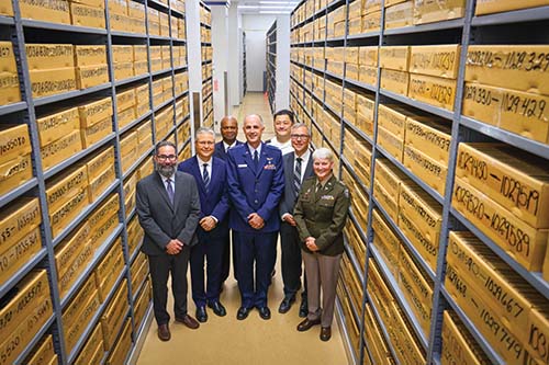
Now, AI is justifying the decision to preserve. The repository’s 55 to 60 million glass slides are being digitized at the rate of 1.5 million slides per year. Increasing numbers of the estimated 32 million formalin-fixed, paraffin-embedded tissue blocks are being examined, and the collection is being curated into unique pathology data sets. Aaron Auerbach, MD, MPH, is the JPC’s director of research, repository, and education.
The JPC’s repository, in addition to being the largest, has advantages over other repositories. “We have drawn and continue to draw specimens from the whole country and internationally as well,” Dr. Lyman says. “We have, in many respects, the best repository for data creation because of the diversity of our samples and the quality of the diagnoses that were originally made, many by pathologists who were leaders in their field.”
Potentially, he says, the JPC could provide a standardized data set for measuring, validating, and judging other algorithms.
The AI-trained computer can accomplish feats “in all kinds of amazing ways,” Dr. Lyman notes. For example, “When we place an immunohistochemical stain on tissue, we can tell if it has a given antigen. But the AI-trained computer can recognize even before staining if the tissue will light up with an immunohistochemical stain or not. It can see morphologic changes that we literally cannot see.” In fact, “It can create an image of the stain before we actually perform the stain and accurately predict what it’s going to look like.”
To enable this, “We need the best quality data to inform our algorithm. The better the data, the better the algorithm.”
A notable example of how algorithms are being informed are the Entamoeba histolytica cases from the JPC collection, now being used to train an AI algorithm to identify these organisms quickly.
“Entamoeba histolytica is a parasite that looks, to the human eye, like human inflammatory cells,” Dr. Lyman says. “But the computer doesn’t become confused. The algorithm creates a computer-generated heatmap that highlights the organisms. What we might have thought was a macrophage is revealed to be the amoeba. It’s especially beneficial to have this kind of application in infectious disease cases where pathologists shoulder a difficult role, akin to looking for a needle in a haystack, and the needle looks like the hay.”
The JPC’s priority is to make an impact on diseases that affect the readiness of active-duty service members and veteran health, says CAP governor Joel Moncur, MD, PhD, MS, deputy director and chief medical officer of the JPC and its former director. “So we look for any and all forms of data to help us understand what those diseases are, and we also apply our personal experience as pathologists.” Algorithms to provide clinical decision support for testicular cancer, the most prevalent cancer among active-duty service members, are one objective. Algorithms to provide early detection of infectious disease are another. “We developed a prototype algorithm that can identify and then classify infectious organisms. We’re excited about the possibility of pathologists recognizing they have something that looks infectious, and by applying this algorithm, it will give them a heatmap to show them where they should look. Then the algorithm will suggest a diagnosis.”
The aim is shorter turnaround time and correct classification, Dr. Moncur notes. “Imagine a military unit in the field in a foreign nation and it has been struck by an infectious disease and the whole unit is taken out of commission. Maybe it’s contaminated water, or an insect or tick bite, or different things that could transmit disease.” Making a diagnosis onsite and getting the right preventive treatment underway is essential, he adds. “Packaging up and shipping a sample that could arrive in two weeks while something is taking out soldiers and sailors and airmen in a matter of hours is haunting.” Using AI and telepathology to get that answer to them much more rapidly, he says, could have an enormous impact.
“These algorithms are kind of like having an AFIP or a JPC pathologist look over your shoulder at what you’re doing and point things out and say, ‘Look here, look there. I think it’s this. There’s a 90 percent likelihood that you’re dealing with Entamoeba histolytica in this specimen.’ That’s the kind of help we want to put in the hands of our military pathologists and the general pathologists in the civilian world.”
Nearing practical application now are prostate cancer algorithms, developed at the JPC with AI/machine learning.
“We have done extensive digitization of prostatectomy specimens,” Dr. Lyman says, “scanning over 2,000 whole-mount prostatectomies,” through the work of pathologist Isabell Sesterhenn, MD, who has curated 100 percent of the prostate cancer cases in the collection. “She personally analyzed each of the whole-mount specimens and circled the areas of prostate cancer, making it easier to train machine learning algorithms.”
The JPC partnered with a data team from the National Institutes of Health and used the large amount of data Dr. Sesterhenn’s work has generated to create algorithms that have been shown to be accurate at diagnosing and grading. The training data included clinical outcomes, Dr. Lyman says, so the algorithm also performs well in predicting prognosis.
None of that is in clinical practice yet. “It’s going through additional validation processes and generalizability testing, and it is potentially limited because it was developed from prostatectomy specimens,” he says. “We don’t yet know how these algorithms may perform on core needle biopsies. Can we use the algorithms on needle cores or only on prostatectomy specimens? This is one area the digital transformation is pushing us to think through.” But, he adds, “We do believe the algorithms will improve efficiency and accuracy in decision-making for practicing pathologists and urologists.”
Soon, clinicians or institutions will be able to approach the JPC with a set of suspected prostate cancer cases and say, “‘Using your AI-created algorithms for support, please provide the diagnosis, grade, and prognosis for these cases.’ We see potential gains for the patients not just in efficiency and accuracy, but eventually in cost savings.”
They expect these algorithms to be ready for clinical use within 12 to 18 months, he estimates.
Proteomics is a relatively new way to look at tissue, Dr. Lyman notes.
“Proteomics is the idea that if we chemically break down tissue in a specific way, we obtain a profile of the proteins in that tissue for that tumor type. It’s something like microscopic fingerprints.”
With 30 to 35 million blocks available, he continues, “It’s a unique privilege and stewardship to be able to access human pathology tissue, mostly from veterans, going back over 100 years, and learn the proteomic profile of a specific disease biopsied from a WWI veteran in 1921,” for example.
“In the digital transformation we’re undergoing,” Dr. Moncur says, “our focus is to make whole slide images from the H&E-stained slides, but it’s important to recognize that behind the majority of those slides is a tissue block. And that is an incredible resource for all sorts of molecular research.”
Among the countless examples of how the repository has been used for that purpose, “the most famous one is the study from the late 1990s on autopsy material from the 1918 Spanish flu pandemic,” Dr. Moncur says. “There were numerous examples of patients from that pandemic in our archives.” AFIP scientists led by Jeffery Taubenberger, MD, PhD, sequenced the RNA genome of the virus, “and were able to basically rewrite the history books,” Dr. Moncur says. They determined it was an H1N1 influenza A virus and eventually determined the full genetic code of that virus so it could be studied in a controlled environment. This finding, he says, turned out to be useful in the COVID-19 pandemic.
“This discovery was followed by advancements in technology, disease surveillance, treatment, and vaccines that were key to the response to the COVID-19 pandemic. And that’s one very nice example of the power of having a very old repository of diseases.” But it’s not the only one. Other disease discoveries made possible by the JPC repository include Reye syndrome, recurrent respiratory papillomatosis, and SMARCB1-deficient renal medullary carcinoma. “That’s in part because not only do we have the slides but we also have the tissue blocks.”
“All across the spectrum of what we do, there are transformative capabilities within reach, some of which have already been developed,” Dr. Moncur says. “Our machine learning algorithm for prostate cancer is a nice example of a unique capability that is not available by any other means to get the most accurate prediction of prognosis for a prostate cancer patient.”
Matching tissue samples with metadata is an important mission of the JPC’s repository, Dr. Lyman says.
“The federal government has extensive high-quality data in its electronic health record, but it’s not always easy to access, and for good reasons. There are important mechanisms and protocols to ensure patient privacy and military security. Because of the federal electronic heath record, we have potential opportunities to link extensive clinical metadata to the JPC tissue repository.”
The system can match the metadata to individual cases without violating privacy protections, he adds. “We are intensely focused on the ethical use of data because we want to ensure veterans support the way their data is being used, that it remains secure and protected and brings significant added value to veterans and the federal government.”
Soft tissue tumors are another focus of the JPC’s data set development. For several years, retired pathologist John Fetsch, MD, who diagnosed soft tissue neoplasms throughout his career, has been curating cases in the JPC repository, Dr. Moncur says. “He has curated over 20,000 cases that we have digitized and collected metadata on. The cases represent every known entity in the World Health Organization classification right now and the full morphologic spectrum of each disease,” he says, “and they form a one-of-a-kind collection of data ready to be applied to support machine learning and develop useful algorithms.”
Dr. Fetsch is now curating soft tissue cases from the JPC repository full time. “Many are decades old,” Dr. Lyman says. “He ensures the diagnosis terminology is up to date and accurate.” It’s labor-intensive but crucial work, he adds. “We expect these efforts to yield far-reaching benefits in the development of powerful AI algorithms.”
Dr. Fetsch is working also to associate the proper metadata with all these cases, and the metadata includes more than patient demographics. For example, Dr. Lyman says, for a skin biopsy diagnosed as melanoma, the metadata might include that the tissue came from a white male who was 56 years old and who died of melanoma 10 years later. “The best metadata includes more than a diagnosis of melanoma. It will also include the depth of invasion, the specific tumor subtype, and, from a veteran perspective, if he had been deployed, for example. We might be able to add that he was treated with immunotherapy and that he recovered. Any relevant data we can include improves the algorithm. In this way, the associated metadata is rarely considered to be complete.”
Dr. Moncur calls artificial intelligence “a team sport,” one that “requires a broad group of experts all working in sync to make something effective and to avoid unintentional harm.”
“There’s a lot of excitement, justifiably, about what we can accomplish technologically,” he says, “but there may not have been as much consideration about avoiding unintentional harm, relating to patient privacy and bias and things like that.” Dr. Moncur has been heartened to see the focus in the past couple of years on the ethics of AI and ensuring a commitment to serving patients while protecting their privacy.
There is also the question of profitability, Dr. Lyman notes. “It’s hard to know how to federate data, to share data and make it profitable for trusted partners assisting in key ways, while keeping the data protected, secure, and private as required by policy and for ethical reasons. These are data governance-type issues, and we are actively working hard to get it right.”
Dr. Moncur sees the pathology field as perfectly suited to develop and use AI, because of its focus on quality management systems and validation and all it does to ensure high-quality work.
“It’s going to surprise people, the positive ways it will become part of their practice, how quickly it comes, and how big a difference it will make,” he says.
The implications for pathology of the expansion in the use of AI are not fully known, Dr. Moncur says, but he is of the view they will be positive.
“At least over the next 10 years, I would anticipate we are all going to become experts in applying the right model under the right circumstances. So our practice is going to change. It doesn’t necessarily mean we’re going to be replaced, but we have the potential to become more efficient and have unique capabilities.”
In addition to the technical and diagnostic expertise pathologists have, “we will become experts in applying machine learning models,” Dr. Moncur says, “just as we would apply another laboratory test.”
Anne Paxton is a writer and attorney in Seattle.