

Amy Carpenter
August 2026—Recognizing and reporting histologic subtypes and divergent differentiation in urothelial carcinoma is critical for diagnosis, prognosis, and treatment.
“Ideally, you want to mention the subtype histologies in pathology reports and quantify the amount,” said Francesca Khani, MD, associate professor in the Department of Pathology and Laboratory Medicine and in the Department of Urology, Weill Cornell Medicine. In a CAP25 session last fall, Dr. Khani spoke of the mimics of benign entities and of other malignancies.
Her first case, illustrative of the importance of urothelial carcinoma subtype, was that of a 73-year-old woman who was referred to urology for lower urinary tract symptoms. On cystoscopy, the urologist noted a cobblestone appearance of the entire bladder mucosa with no discrete lesions identified. “They presumed clinically that this was cystitis cystica et glandularis—didn’t even biopsy it,” Dr. Khani said. The patient was given multiple antibiotic regimens for a urinary tract infection, but the symptoms persisted.
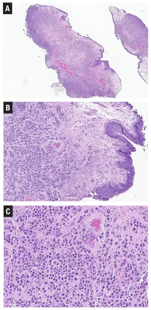
The patient had a repeat cystoscopy and then a transurethral resection, in which a dense infiltrate was seen in the lamina propria; at low magnification it resembled a chronic inflammatory process (Fig. 1A).
At higher magnification (Fig. 1B), the cells in the lamina propria were seen to be more infiltrative and have a discohesive growth pattern. “They show single dispersed cells and some linear arrangements of the cells,” Dr. Khani said, adding that the overlying urothelium notably was benign. At even higher magnification (Fig. 1C), the cells “exhibit a plasmacytoid appearance with an eccentrically placed nucleus, so they look a lot like plasma cells.”
Plasmacytoid urothelial carcinoma comes to mind, she said, and is the top of the differential. “But in a case like this where you don’t have an in situ component, you want to rule out other malignancies, especially because it doesn’t have a classic, conventional-appearing urothelial carcinoma component.” Also in the differential: metastatic carcinoma—lobular breast carcinoma or a signet ring/diffuse-type GI primary; lymphoma/plasmacytoma; an odd-looking sarcoma; and melanoma.
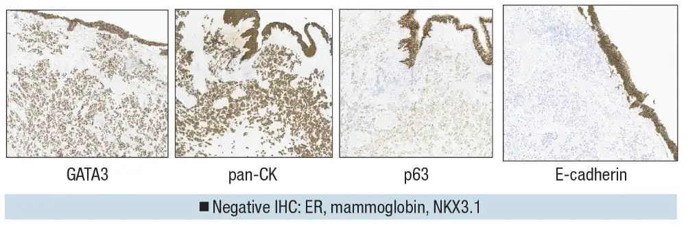
GATA3 and pan-cytokeratin were diffusely positive (Fig. 2), which points toward a carcinoma, likely either breast or urothelial, considering this morphology. This immunoprofile points away from it being lymphoma or sarcoma or anything else that would be pan-CK negative. Additionally, p63 was weakly but still diffusely positive in the tumor. The p63 positivity helped to rule out lobular breast cancer, she noted, which is usually p63 negative. E-cadherin was also negative, with loss of membranous staining in the tumor cells. This would be seen in a lobular breast cancer, but plasmacytoid urothelial carcinoma can also show loss of E-cadherin, as in this case.
In looking further at the breast carcinoma differential, estrogen receptor and mammaglobin can be used. “ER can sometimes be positive in urothelial carcinoma but would usually have weaker staining,” Dr. Khani said, “whereas a lobular breast cancer, lower-grade looking like this, would usually be more diffusely positive.”
NKX3.1 is a very sensitive and specific marker for prostate cancer, “so why use it in a woman?” she asked. “A lot of pleomorphic lobular carcinomas have been shown to be NKX3.1 positive, so you can use that marker to help rule it out,” particularly if a metastatic pleomorphic lobular breast cancer is in the differential.
With the GATA3, p63, and pan-CK positivity, she said, the diagnosis is plasmacytoid urothelial carcinoma.
Divergent differentiation includes urothelial carcinomas that have squamous, glandular, or trophoblastic differentiation. Urothelial carcinoma histologic subtypes consist of nested, large nested, tubular and microcystic, micropapillary, lymphoepithelioma-like, plasmacytoid, giant cell, lipid-rich, clear cell (glycogen-rich), sarcomatoid, and poorly differentiated. The 2022 World Health Organization classification of urinary tract tumors moved to the use of subtypes in invasive urothelial carcinoma from the prior use of variant histology to avoid confusion with a molecular alteration, Dr. Khani said.
The difference between divergent differentiation and subtype can be a “little semantic,” she said, citing an editorial, “Urothelial Carcinoma—Are We Moving Beyond ‘Divergent Differentiation’ and ‘Histologic Subtype’?” (Wobker SE, et al. Am J Clin Pathol. 2025;164[2]:141–144). Often both are present in the same tumor. “You can have a urothelial carcinoma that has some squamous differentiation. You can also have another area that has some subtype histology,” Dr. Khani said.
Wobker and Smith, in their editorial, write that divergent differentiation is the identification of tumor cells transdifferentiating toward features of cells of a nonurothelial origin, whereas subtypes represent urothelial cells that have taken on a nonconventional, distinctive architectural growth pattern, nonetheless retaining their urothelial cytologic and immunohistochemical features. “However, this isn’t always true,” Dr. Khani said, and as the authors noted. Some subtypes, such as sarcomatoid, represent a differentiation of the urothelial phenotype. “So there’s a lot of overlap in the terminology. Nonetheless, the WHO classifies these as separate entities.”
The 2022 WHO classification of urinary tract tumors is largely unchanged from the 2016 edition except for the modification of two histologic subtypes, she said. The large nested and nested subtypes are now two separate subtypes based on nest size, and microcystic urothelial carcinoma is now tubular and microcystic to reflect the architectural pattern.
The nested, large nested, and tubular and microcystic urothelial carcinoma subtypes mimic benign entities, Dr. Khani said, and the plasmacytoid, lipid-rich, and clear cell (glycogen-rich) subtypes mimic other malignancies. “Almost all of these [subtypes] can mimic other malignancies, so you always want to rule out malignancies from other organ systems.”
The micropapillary, plasmacytoid, giant cell, sarcomatoid, and poorly differentiated urothelial carcinoma subtypes have the strongest evidence of more aggressive behavior, she said, “even when you control for the stage of a tumor.” The most common of the five are micropapillary, plasmacytoid, and sarcomatoid subtypes. The lymphoepithelioma-like urothelial carcinoma subtype, which responds well to therapy, is the only subtype with a more favorable prognosis.
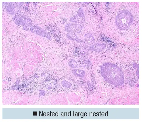
The nested and large nested variants of urothelial carcinoma are composed of small or large nests of cells that infiltrate the lamina propria. They are often muscle invasive and typically have “a very infiltrative growth pattern but the cytology is usually quite bland,” Dr. Khani said.
Taken in isolation, the large nests seen in Fig. 3 could mimic a florid proliferation of von Brunn nests, especially if they are superficially located. “Being cytologically bland, this can be a very difficult distinction, and that infiltrative growth pattern is most helpful,” particularly when the nests are seen invading muscularis propria, as in this case.
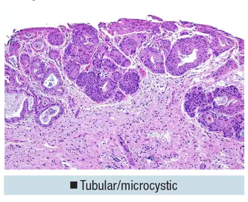
The tubular and microcystic subtype typically has fairly bland histology and glandular, tubular structures that mimic cystitis cystica et glandularis (Fig. 4). “A florid proliferation of cystitis cystica can look a little similar to the tubular and microcystic pattern.”
In Fig. 5A is a tumor that was in the upper urothelial tract. Nests are in the lamina propria, and it is difficult to tell at low magnification if the tumor is a florid proliferation of von Brunn nests, she said, or an invasive nested subtype of urothelial carcinoma.
A small nest below the muscularis layer in the muscularis propria is seen in Fig. 5B. “Make sure you always, in these resections, look at not just the surface,” Dr. Khani said, “and if you’re struggling with what’s at the surface, sometimes looking below the surface for more invasion can be helpful.” Doing so helped determine that the rest of this tumor was an invasive pattern.
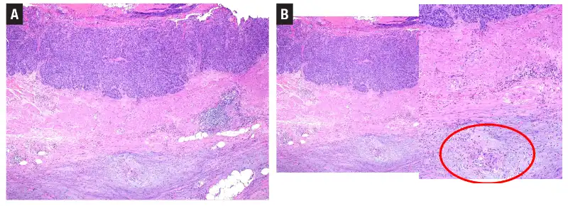
Thus, the histologic features that distinguish subtypes from benign entities are as follows:
- In the nested and large nested subtypes, look for an irregular/infiltrative border. Typically, florid proliferation of von Brunn nests will have a more well-defined border. “If you see nests within or beyond the muscularis propria that are truly invasive, that would help with the diagnosis,” Dr. Khani said.
- In the tubular and microcystic subtype, the usual features of invasion—paradoxical differentiation and single cells in the lamina propria—are often useful. “Typically, the size of the nests in the tubular structures are a little more variable than they are in benign entities, like a cystitis cystica.”
This can be challenging on very small cystoscopic biopsies, she said, where little is seen below the layer of the lamina propria or if it is difficult to determine whether there is an infiltrative border.
Her practice tip: If suspicious for a nested subtype but difficult to determine on a small biopsy, “say so and recommend more sampling.” A sample top-line diagnosis could be: atypical nested urothelial proliferation. A note can reflect the differential diagnosis and level of suspicion and recommend the additional sampling if clinically indicated.
Turning to subtypes that mimic nonurothelial malignancies, Dr. Khani said the clear cell (glycogen-rich) subtype is composed of sheets of clear cells with well-defined membranes and clear cytoplasm, owing to the abundance of glycogen (Fig. 6). This subtype looks similar to a renal cell carcinoma; it should be distinguished from that as well as from clear cell adenocarcinoma, both of which would be PAX-8 positive. A clear cell (glycogen-rich) urothelial carcinoma should be diffusely GATA3 positive.
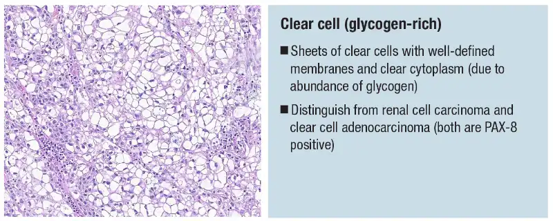
If something that looks like a clear cell (glycogen-rich) urothelial carcinoma is seen in a lymph node, “you wouldn’t jump to urothelial carcinoma as being the primary,” so it is important to mention these in a primary section, Dr. Khani said. “If it spreads later on, it helps a pathologist in the future recognize it could be urothelial when it doesn’t immediately look like that.”
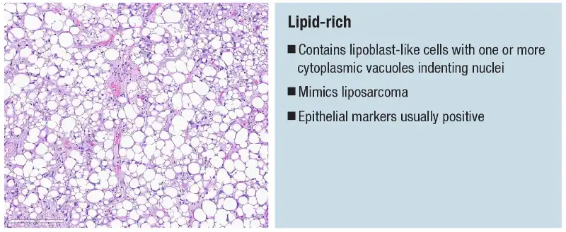
The lipid-rich subtype also mimics nonurothelial malignancies (Fig. 7). It’s a urothelial carcinoma that contains lipoblast-like cells with one or more cytoplasmic vacuoles indenting the nuclei. It mimics a liposarcoma; however, epithelial/urothelial markers are usually positive, she said.
Subtype histologies are rarely seen in the pure form, she said. “Typically, you will see another conventional component and that’s always helpful. But if you do see isolated histology [as seen in Fig. 7], it’s always good to rule out other malignancies.”
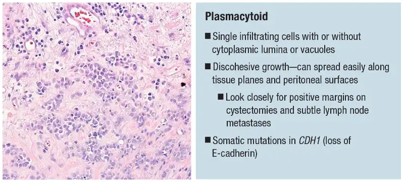
The plasmacytoid subtype (seen in the case reported earlier) typically has single infiltrating cells with or without cytoplasmic lumina or vacuoles and with discohesive growth (Fig. 8). “These tumors can spread easily along tissue planes and peritoneal surfaces,” Dr. Khani said, advising a close look for positive margins in cystectomy specimens. “The lymph node metastases can be very subtle because of the cytology of these cells.” Plasmacytoid urothelial carcinomas are often associated molecularly with mutations in CDH1 (loss of E-cadherin).
A variety of morphologies can show similarly discohesive growth patterns, she said, and thus there is some interobserver variability. E-cadherin IHC can help, but E-cadherin loss is seen in only about 74 percent of plasmacytoid urothelial carcinoma cases and is sometimes subtle. “I don’t routinely stain anything that looks plasmacytoid with E-cadherin, but if on the fence, it can maybe help push you one way or the other.”
Dr. Khani’s second case was that of a 76-year-old man with a bladder tumor and a recent urine cytology positive for high-grade urothelial carcinoma. In the transurethral resection (Fig. 9A) is a nested proliferation, with possibly inverted growth and solid sheets of cells, as well as a hint of papillary architecture with the fibrovascular cores (Fig. 9B).
In other areas of the heterogeneous tumor were nests of cells that prompted consideration of a nested urothelial carcinoma (Fig. 9C). These are areas that have more of a discohesive growth pattern, with single cells spreading all over. A single cell infiltrative pattern within the muscularis propria invading the muscle is seen (Fig. 9D).
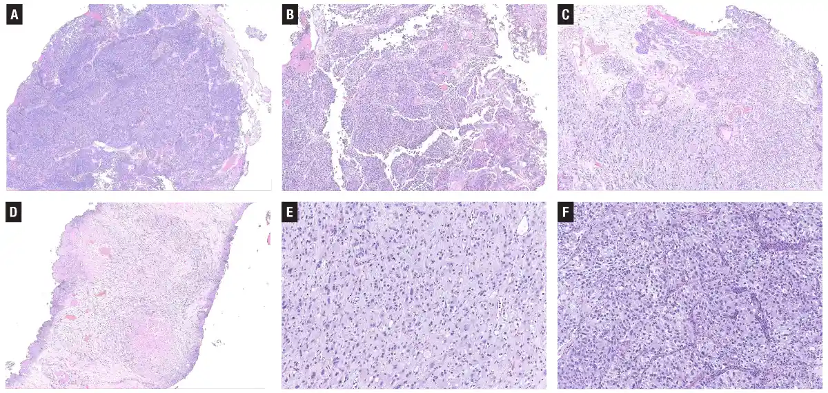
Some cells have a somewhat eccentric nucleus with eosinophilic cytoplasm and discohesive-type growth (Fig. 9E), and other areas have cytoplasmic clearing and a clear cell appearance (Fig. 9F). “So a lot of different looks in this one tumor.” The heterogeneity brought to mind urothelial carcinoma.
Some areas of the tumor have very prominent nucleoli (Fig. 10), and the cytoplasmic clearing looks a little different from a real clear cell (glycogen-rich) carcinoma, “so I was suspicious that maybe we should be thinking about other things.”
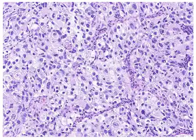
Some areas are slightly more paucicellular. The nuclei are cytologically bland and have some cytoplasmic clearing. “Then when I reviewed this case more thoroughly,” she said, “I thought there wasn’t a good, definite in situ urothelial carcinoma component.”
Immunostaining found the entire tumor to be strongly positive for NKX3.1 in all areas. “The solid areas that I thought might have been a little nested-appearing were negative for NKX3.1, and they were all well circumscribed at the surface and positive for GATA3,” Dr. Khani said. The solid areas were von Brunn nests mixed into the surface of the tumor, “but the rest of it was prostatic adenocarcinoma.”
“Focally, some of the changes were seen because the patient had had hormone therapy treatment for prostate cancer, and this pertinent history was not provided to us initially.” Malignant cells were seen in the urine (recall the positive urine cytology), but they were not a high-grade urothelial carcinoma. “It’s always good, in a case like this, to alert your cytopathologist that they might not be dealing with a urothelial primary.”
The lesson: Always consider other primary malignancies, especially in the absence of in situ urothelial component or if the morphology seems unusual for any subtype, even if that unusual morphology is very focal, she said. “It’s worth working up with immunostains if there’s anything that isn’t quite perfect for urothelial carcinoma. The treatments are very different.”
Dr. Khani turned to the prognostic and therapeutic implications of urothelial carcinoma subtype histologies.
For muscle-invasive disease, the prognosis in pT2–T4 is influenced not only by pathological stage at cystectomy but also the presence of aggressive urothelial carcinoma subtypes: plasmacytoid, micropapillary, neuroendocrine/small cell, and sarcomatoid.
Retrospective data show limited benefit to neoadjuvant chemotherapy, particularly in the micropapillary and sarcomatoid subtypes. “But neoadjuvant chemotherapy is still recommended for all subtype histologies.”
The treatment implications for the aggressive subtypes are more important in the nonmuscle-invasive setting, Dr. Khani said, because in the National Comprehensive Cancer Network guidelines (version 1.2025), subtype histology puts patients in the American Urological Association risk stratification high-risk category. Subtype presence is also considered a very high-risk feature.
“And the presence of a very high-risk feature in a pT1 bladder cancer warrants the consideration of cystectomy,” she said, noting it’s listed as the preferred primary treatment. “Diagnosing these subtypes is getting even more important in the more minimally invasive setting of T1 disease.”
Some physicians treat nonmuscle-invasive bladder cancer cases more conservatively, she said, especially if there is a very small focus of invasion versus a larger area of T1. “Nonetheless, you could be buying this patient a very radical surgery for calling out an aggressive subtype.”
Giant cell urothelial carcinoma, a rare aggressive subtype, has pleomorphic giant cells, and the poorly differentiated subtype, also aggressive and rare, lacks morphologic features of urothelial origin.
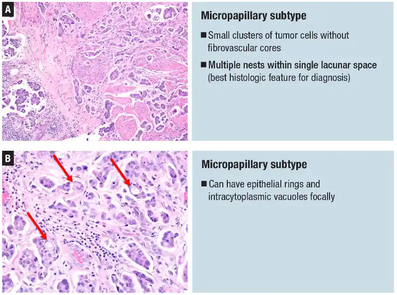
The micropapillary and sarcomatoid aggressive subtypes are fairly common. Small clusters of tumor cells without fibrovascular cores are seen in the micropapillary subtype (Fig. 11A). Multiple nests are seen within single lacunar spaces, “and that’s the best histologic feature that’s most reproducible for diagnosis,” she said, “because this can mimic lymphovascular invasion and retraction artifact that you tend to see in invasive urothelial carcinomas” (Sangoi AR, et al. Am J Surg Pathol. 2010;34[9]:1367–1376). Areas of lymphovascular invasion are seen often in aggressive cases, “so you might see both.”
Epithelial rings and intracytoplasmic vacuoles focally are other features that can be helpful in identifying the micropapillary subtype (Fig. 11B). This subtype also often shows HER2 overexpression. (A CAP biomarker project team, composed of members of five CAP committees—Cancer, Pathology Electronic Reporting, Immunohistochemistry, Center Guideline, and Molecular Oncology—issued a guidebook to HER2 testing in solid tumors, including the bladder, published online June 19, 2026 in Archives of Pathology & Laboratory Medicine. doi:10.5858/arpa.2025–0593-CP.)
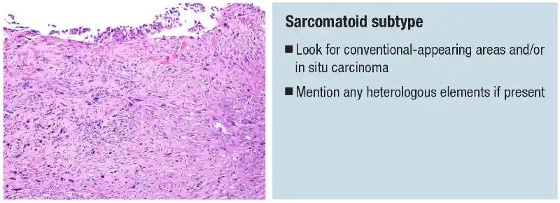
If the tumor has a pure sarcomatoid appearance with spindled pleomorphic cells, “you want to consider a very broad differential diagnosis,” Dr. Khani said. Immunohistochemistry can be helpful. “But they may lose staining for urothelial markers GATA3, high-molecular-weight cytokeratin, and p63. They’re worth a shot anyway,” she said, “because sometimes there are areas that retain those markers.”
Looking for a conventional component and/or carcinoma in situ can help determine whether a tumor is truly a sarcomatoid subtype. In the case in Fig. 12, there was CIS at the surface and more epithelioid-looking areas in the invasive component, Dr. Khani said. Heterologous elements should be reported if present.
A lymphoepithelioma-like urothelial carcinoma will have sheets and clusters of undifferentiated cells with poorly defined cytoplasmic borders and a syncytial appearance. The tumor cells have prominent nucleoli and are associated with dense, chronic inflammatory infiltrate. They’re highly sensitive to chemotherapy and enriched for PD-L1 expression, showing a good response to immune checkpoint inhibitors.
Metastatic sites often reflect subtype histology, so reporting the subtype histology provides information for subsequent encounters. Reporting the percentage of subtype histology within a sample is also recommended, but there is no recommended percentage for calling a pure subtype.
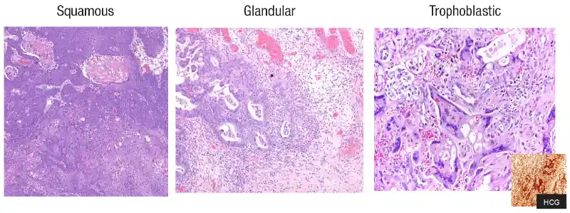
Divergent differentiation (Fig. 13)also has diagnostic and prognostic therapeutic implications. It’s important to distinguish squamous and glandular differentiation from a pure squamous or a pure adenocarcinoma of the bladder, which are considered nonurothelial tumors. To distinguish between them, Dr. Khani said, look for a conventional urothelial component (Fig. 14).
A pure squamous carcinoma is considered a nonurothelial diagnosis and is more common in Egypt and other African countries owing to its association with schistosomiasis. It can be more commonly associated with chronic bacterial infections in neurogenic bladders.
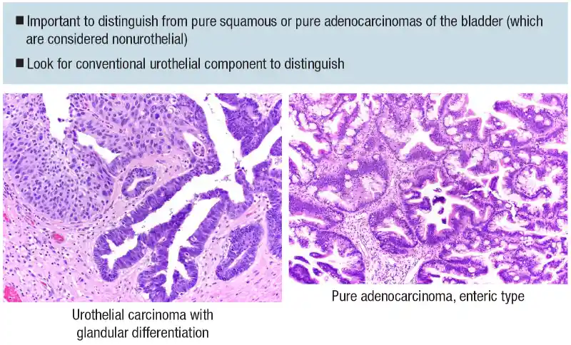
When pure squamous carcinoma is seen in women, cervical squamous carcinoma with secondary bladder involvement is an important differential diagnosis. HPV RNA in situ hybridization can be helpful, and gynecologic clinical history is most important to the diagnosis, Dr. Khani said (Schwartz LE, et al. Am J Surg Pathol. 2016;40[1]:27–35). “We often don’t think about cervical primaries now because the incidence has gone down with the HPV vaccine, but that’s more reason to think of it.”
Pure adenocarcinomas can have enteric (histologically indistinguishable from GI adenocarcinoma), mucinous, or mixed morphology. Immunostains (CDX2+/CK20+) are not helpful because they will pick up expression seen in intestinal primaries, and clinical correlation is necessary for definitive diagnosis.
There is no proven role for neoadjuvant or adjuvant chemotherapy in pure squamous or adenocarcinoma of the bladder, Dr. Khani said, so distinguishing these from divergent differentiation is critical for treatment.
Dr. Khani shared her final case: A 77-year-old woman was found to have a 1.0-cm enhancing mass in the dome of the bladder (an incidental finding seen on imaging). A transurethral resection was performed.
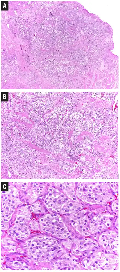
The tumor (Fig. 15A) shows a very infiltrative pattern, a nested architecture, and appears to be invading muscle. At higher magnification (Fig. 15B), it appears sheet-like, with a nested appearance, though not well-defined nests. The tumor seen in Fig. 15C has a bit of a neuroendocrine look with salt-and-pepper chromatin to the cells and amphophilic cytoplasm.
There was no in situ component, and the immunoprofile was consistent with a urothelial carcinoma with neuroendocrine differentiation. GATA3 and synaptophysin were strongly positive, “giving mixed signals,” Dr. Khani said. “It doesn’t seem right. We have nested architecture and it’s cytologically bland, but we have a neuroendocrine marker that’s very positive.”
The diagnosis: paraganglioma. “If you’re considering a paraganglioma, you should always throw on a cytokeratin because paragangliomas are positive for GATA3 and you can go down the tubes thinking it’s a urothelial carcinoma,” she said. “But in this case, pan-cytokeratin and p63 were completely negative.”
Paraganglioma is another mimicker of urothelial subtypes, she said, particularly the nested variant. “They can show deep invasion, but they don’t have any histologic criteria for malignancy, typically.”
Paragangliomas usually are more bland than a typical urothelial carcinoma that has divergent differentiation to neuroendocrine, but they nonetheless have a neuroendocrine phenotype. GATA3 positivity in paragangliomas represents a pitfall for urothelial carcinoma.
Amy Carpenter is CAP TODAY senior editor.