

Amy Carpenter
September 2026—Amyloidosis has become a treatable disease—if the fibril type is identified accurately and quickly.
That and the variability seen in its workup and diagnosis were the impetus for the recently published CAP amyloidosis guideline (Miller DV, et al. Arch Pathol Lab Med. 2026;150[3]:179–193).
The co-chairs of the guideline expert panel provided, in a CAP25 session, what they described as their own tips and tricks for amyloid detection and fibril typing, reported here.
There are at least 42 amyloid subtypes, said panel co-chair Billie S. Fyfe, MD, professor in the Department of Pathology and Laboratory Medicine at Rutgers Health Robert Wood Johnson Medical School. “And with mass spectrometry, more amyloid types are being identified each year.”
The fibril type dictates the treatment, said co-chair Dylan V. Miller, MD, professor of pathology at the University of Utah School of Medicine and chair of the CAP Immunohistochemistry Committee. “In my mind, that sounds a lot like a predictive marker test,” he said, “like PD-L1 and HER2 and others.”
If left untreated, 25 percent of patients with immunoglobulin light chain (AL) amyloidosis die within six months of diagnosis, and a similar percentage with amyloid transthyretin (ATTR) amyloidosis die within 24 months of diagnosis. All forms of amyloidosis are underdiagnosed (Gertz MA, et al. JAMA. 2020;324[1]:79–89).
Now to their tips and tricks, but first a recap of the guideline recommendations, or, as Dr. Miller puts it, the “guideposts.”
The first recommendation says in patients with suspected systemic amyloidosis, pathologists may screen cytology specimens (conventional smears and/or cell blocks) of aspirated abdominal fat for detection of amyloid. The recommendation notes that individual laboratories should determine and optimize the best preparation methods and that ancillary testing techniques should be validated on cytologic material. If cytologic smears only are prepared in the absence of a cell block, it says, the ability for further testing, including subtyping, is limited.
There are data to support the screening of cytology specimens by conventional smears or cell blocks, Dr. Miller said. “If you’re not using a tissue-based method, you should be sure the method you’re using works for detecting amyloid.” If using an air-dried smear, “you should know when you do Congo red on that smear you are able to detect amyloid in your lab.” And if using a limited sample like an air-dried smear, he added, the fibril typing will not be able to be performed. “There may be a need to get more tissue for doing that important part of the diagnostic workup.”
Recommendation No. 2 says when evaluating specimens for the presence of amyloid, pathologists should use the Congo red staining method. Other methods can be used but should be validated against Congo red or electron microscopy and must show equivalency.
The many published articles about Congo red and the few articles about any other amyloid stain explain the lack of evidence supporting a recommendation for other stains, Dr. Miller said. However, many laboratories are successfully using sulfate Alcian blue, thioflavin T, and other stains appropriate for amyloid detection. “We didn’t want to discourage them from continuing that practice,” he said, provided the laboratory validates the alternative staining method.
Recommendation No. 3 says when assessing Congo red histochemistry, pathologists may add fluorescence microscopy with the tetramethylrhodamine isothiocyanate/Texas red filter to increase sensitivity for amyloid detection.
“You don’t need to buy a fluorescence scope to do this,” Dr. Miller said, “but if you have one, this is a helpful thing you can do to improve sensitivity of your Congo red interpretation.”
The fourth recommendation says in patients with amyloidosis for whom therapy is being considered, mass spectrometry should be used to identify the fibril protein type in order to optimize diagnostic yield and the use of tissue. In renal amyloidosis, it notes, amyloid fibril typing may often be accomplished successfully by immunofluorescence, though reflex to MS-based proteomics should be performed in difficult or equivocal cases.
The three good practice statements in the guideline are as follows:
• When specimens are received for detection of systemic amyloidosis, pathologists should evaluate for the presence of amyloid using validated method(s), and the validated method(s) used should be identified in the pathology report.
For a fat pad or other surrogate biopsy that is negative, Dr. Miller said, “be very clear that that doesn’t exclude the possibility of amyloid in a target organ or elsewhere in the body.”
• If a clinical concern for amyloidosis persists after a negative biopsy from a surrogate site but the potentially affected organ (or organs) was not sampled, then suggesting a biopsy of the potentially affected organ(s) and/or recommending suitable archived specimen(s) to evaluate is appropriate.
“You may need to recommend a biopsy of a potentially affected organ,” he said. Or if there is a tissue sample in the archive from a recent procedure, “you may want to pull that block and perform Congo red on that to see if it may have some amyloid.”
• In patients with amyloidosis who are being considered for therapy, pathologists should determine the fibril protein type.
Among the methods for identifying amyloid fibril is IHC, which can be effective, Dr. Miller said, but the limitations are that the panels are only as good as the optimization of each of the assays in the panel (Gonzalez-Lopez E, et al. J Am Coll Cardiol. 2024;83[11]:1085–1099).
In addition, it’s a “closed-ended exercise,” he said. “You can only interrogate what you have antibodies for, compared with mass spectrometry, which will detect the sequence of any peptide that is in there. It’s more open-ended in terms of its identification approach.”
Another method, immunoelectron microscopy, combines confirming the presence of the definitive amyloid fibrils with the specificity of monoclonal antibodies that have gold attached to them. It’s not only the amyloid fibrils that can be seen but also the specificity of the gold particles binding to the fibrils.
Mass spectrometry can be done on paraffin tissue and is the gold standard, Dr. Miller said, noting it has very high sensitivity and specificity and can detect the amyloid type no matter what the fibril peptide may be, and even multiple peptide types. In rare cases, he said, there is more than one type of amyloid overlapping (Gonzalez-Lopez E, et al. J Am Coll Cardiol. 2024;83[11]:1085–1099).
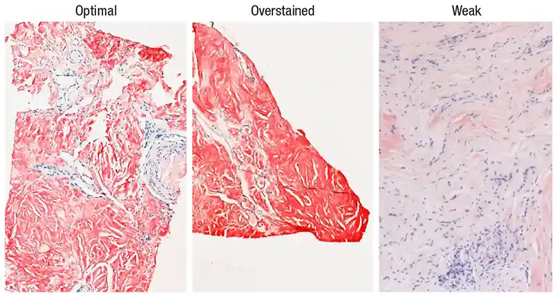
The first of the practical tips and tricks for slide reading when the question is amyloid is to check the on-slide tissue control (Fig. 1). Ideally, it should show good staining of what is supposed to be amyloid and have a negative component, Dr. Miller said, such as the vessel that did not pick up the Congo red stain in Fig. 1 (left image).
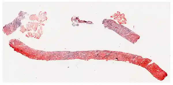
“Not everything that’s red on a Congo red is positive for amyloid,” he said of a case he had a week before his talk (Fig. 2). “This is not immunohistochemistry, it’s not a trichrome. And Congo red tends to be very prone to overstaining and nonspecific staining, so do not just look at a slide at 2× and say it is Congo red positive even though it’s pretty red.”
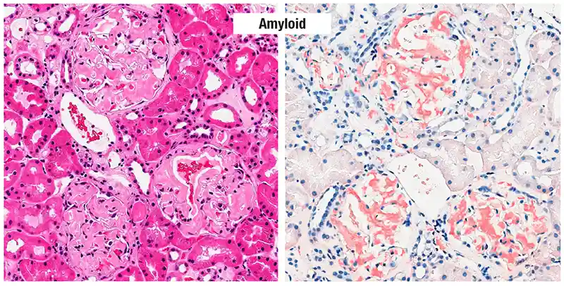
He always begins with the H&E. “For me to call something amyloid, it has to look like amyloid on the H&E,” though that is not sufficient. “I can’t diagnose amyloid on an H&E, but it always has to start there.”
Prototypical renal glomeruli can be seen in Fig. 3 (left). The material should be pink and glasslike, smooth, or waxy. There should be no cells embedded in the material.
“So if the H&E looks like it could be amyloid, and the Congo red is hitting everywhere where it could be amyloid, then I’m pretty suspicious,” he said (Fig. 3, right). “I’m almost there to saying it looks like amyloid.”
By contrast, in Fig. 4 (left), no smooth, glassy material can be seen in the kidney biopsy of a patient with diabetes. Cellularity is visible where there is pink material and there is a bit of fibrosis (left image, lower middle).
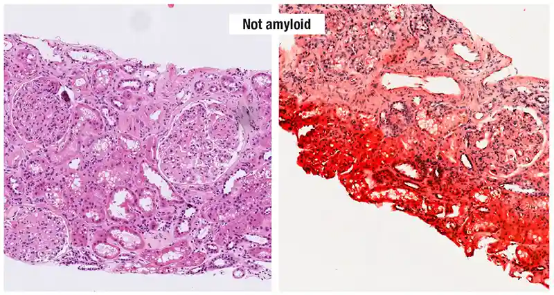
“If we were to go higher in the fibrotic area, we would see not a smooth texture,” he said, but “probably ropy collagen strands or some fine fibrillar material.” Amyloid fibrils can be seen only with electron microscopy. “You shouldn’t see any fibrillar material or texture on the H&E in true amyloid.”
In the image on right, the Congo red is heavily overstained; it is not specific and does not match anything that would look potentially like amyloid on the H&E. Some red staining can be seen in the same image in a vessel, which Dr. Miller looks for because, he said, “amyloid loves to deposit in vessel walls.” But only smooth muscle is seen in the vessel walls in the image on left, and there is no other extracellular matrix material to indicate the possibility of amyloid deposits.
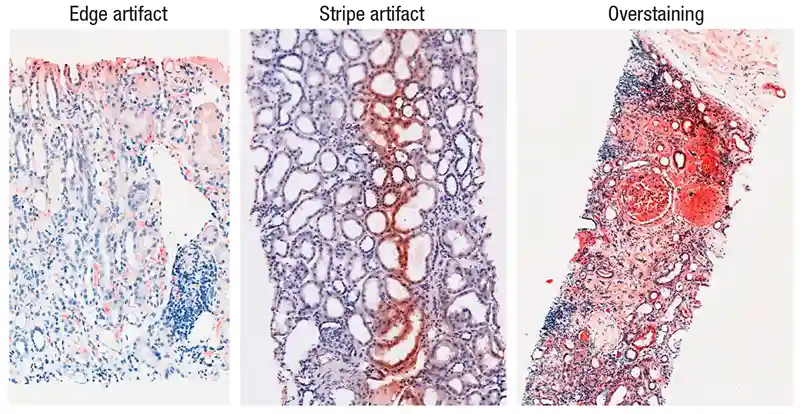
Edge and stripe artifacts and heavy overstaining in a regional pattern are seen in Fig. 5. “Overstaining and artifacts are almost the rule now, at least in my lab,” Dr. Miller said. “We use the Dako [Agilent] Artisan autostainer for Congo red and we’re never going back to hand-staining.”
Nonspecific congophilia is a pitfall to be aware of, he cautioned. It’s a tissue sample that stains red with Congo red but is not amyloid. Chondroid, erythrocytes, skeletal muscle but also smooth muscle, and elastic tissue in vessels tend to show nonspecific congophilia. “Be careful if you’re seeing Congo red in vessels, and it looks a little smooth and amorphous, that it’s not just elastic tissue,” he said. Dense collagen (blue-white birefringence) and fibrin can pick up Congo red nonspecifically.
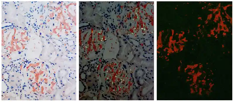
Fluorescence can be helpful in highlighting Congo red positivity. In Fig. 6 polarized light microscopy is in the center; fluorescence is on the right.
“We learned in medical school about apple green birefringence,” Dr. Miller said. “I’m not sure I’ve seen too many apples that are the color I see under polarized light. For me, it’s not so much a Granny Smith apple but more like a Golden Delicious kind of green apple.” The amyloidosis guideline uses the term characteristic birefringence. “That’s preferred in the amyloid nomenclature now because it’s more than green.” Orangish hues can be seen. “These should be specific and match what you’re seeing on the Congo red stain by nonpolarized and polarized microscopy.”
(Click below to see a video, courtesy of Dr. Miller, showing birefringence detection in a tissue sample under polarized light microscopy.)
Cardiologists are now diagnosing transthyretin amyloidosis themselves on the basis of scans, said Dr. Fyfe, who spoke of heart and fat pad biopsies using her cases from Rutgers Health Robert Wood Johnson Medical School. “Amyloid is a disease of the aged, and patients are coming in with an MGUS [monoclonal gammopathy of undetermined significance] and a scan, and the scan might be equivocal,” she said. Heart biopsies are characteristically fairly straightforward, she noted, but “are sometimes now a bit trickier.”
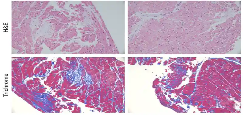
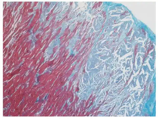
In Fig. 7 are the H&E (“definitely amyloid, maybe subtle”) and trichrome stains of an endomyocardial biopsy from a patient with MGUS. “You can have fibrosis in a heart that has amyloidosis,” Dr. Fyfe noted; the amyloid destroys tissue and scar tissue replaces it. The trichrome looks blue but for amyloid deposits is supposed to look a little bluish-gray. A closer look in Fig. 8 reveals the blue-gray appearance.
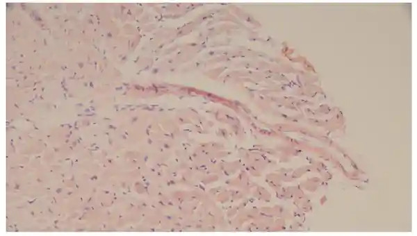
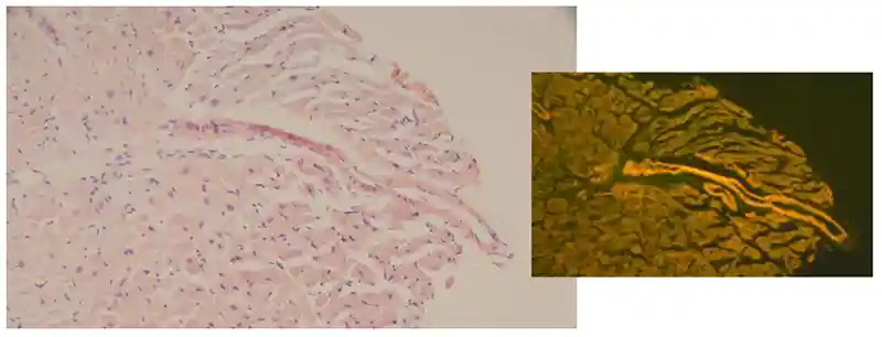
An endomyocardial biopsy from another patient with MGUS shows subtle Congo red staining on the H&E (Fig. 9), but the Congo red fluorescence microscopy (Fig. 10) reveals strong vascular staining. In a case like this, pathologists at Dr. Fyfe’s institution will freeze tissue for additional light chain immunofluorescence staining. “At this time, I don’t rely only on the immunofluorescence for fibril typing; I also send for mass spectrometry,” she said. “But I’m trying to accumulate data on the correlation between fluorescence and mass spectrometry in these sorts of cases.”
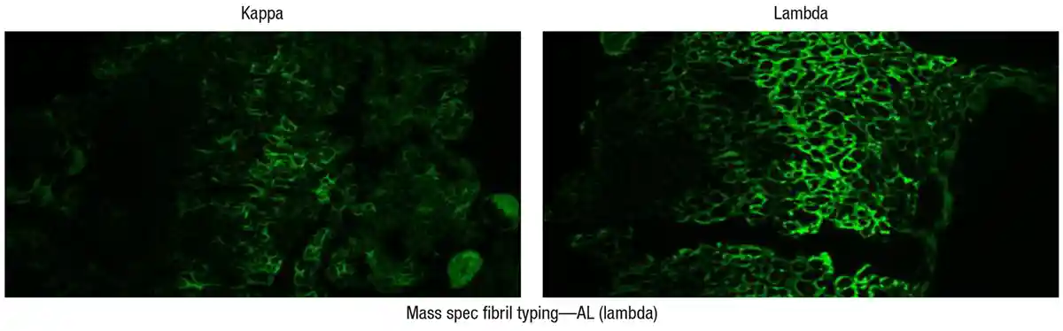
In Fig. 11 is a biopsy from a patient who had a lambda light chain MGUS. The lambda staining on immunofluorescence was positive (right), but there was also kappa staining (left). She ordered mass spectrometry fibril typing, which confirmed the patient had AL (lambda) amyloidosis.
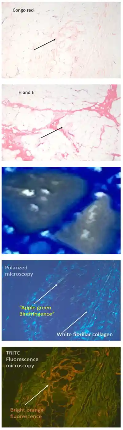
“Fat pads are another thorn in people’s side,” Dr. Fyfe said. In Fig. 12 the Congo red stain is focally positive. White fibrillar collagen is seen under polarized microscopy, as is birefringence. Bright orange fluorescence was observed under fluorescence microscopy. “So it was diagnostic to me,” Dr. Fyfe said. “The patient did go on to get a further workup, and it was an AL amyloidosis.”
Not covered in the guideline is screening for amyloid in nontargeted tissues. “The Venn diagram of amyloidosis overlaps with some other disease conditions significantly,” Dr. Miller said, “and carpal tunnel syndrome and lumbar stenosis happen to be two of those other circles that overlap.”
The guideline does not suggest staining every carpal tunnel or lumbar stenosis specimen with Congo red, but it’s important to have the awareness, he said. “And if a clinician calls and says, ‘I did a carpal tunnel surgery on this person’ or ‘They had it done, and I’m a little worried they might have amyloid,’ they probably have a good point and you probably should do Congo red. There is a reasonable chance you might find something there.”
The guideline addresses only systemic forms of amyloidosis, but localized amyloidosis is seen in a few sites: larynx, skin, bowel (in areas of injury), and the urinary tract, which is rare. “The nuanced twist to be aware of,” he said, “is that you can have localized amyloid deposits where the fibril type is a type that can be systemic. For example, an amyloidoma of the lung may be AL lambda-type amyloid, but it may not mean the patient has systemic amyloidosis. So it’s tricky.”
“Just because you’ve diagnosed a potentially systemic form in a tissue biopsy,” he continued, “doesn’t mean they have the systemic form of that disease.”
If patients make repeated injections into their abdominal fat pad, some of the drugs and agents can misfold and undergo deposition in a manner identical to amyloid deposition. “You may see amyloid in their fat pad biopsies,” Dr. Miller said, “but if they’re a diabetic who’s been injecting insulin for years, be very careful about recognizing this may be in a localized form.”
Asked about seeing amyloid deposition from the injection of GLP-1 agonist-type drugs, he said, it’s “possible but probably needs more time to evolve.”
The therapies for light chain amyloidosis include those that are plasma cell and immune directed: daratumumab (anti-CD38), cyclophosphamide, dexamethasone, and belantamab mafatodin (ADC); proteasome inhibitors (bortezomib); autologous stem cell transplant; and newer anti-amyloid antibodies that bind to fibril epitopes, leading to macrophage activation and dissolution (CAEL-101, birtamimab, AT-e).
The directed therapy for transthyretin amyloidosis is vastly different, Dr. Fyfe said. “Transthyretin is made in the liver, and we can try to inhibit transthyretin production with RNA silencing.” For ATTR, tafamidis is an FDA-approved stabilizer that prevents TTR tetramer dissociation. “And similar to light chain, they’re trying seeding inhibitors [TabFH2] and antibodies [PRX004, NI006] to break up the amyloid fibrils.”
Amy Carpenter is CAP TODAY senior editor.