


CAP TODAY and the Association for Molecular Pathology have teamed up to bring molecular case reports to CAP TODAY readers. AMP members write the reports using clinical cases from their own practices that show molecular testing’s important role in diagnosis, prognosis, and treatment. The following report comes from Columbia University College of Physicians and Surgeons. If you would like to submit a case report, please send an email to the AMP at amp@amp.org. For more information about the AMP and all previously published case reports, visit www.amp.org.
Mine Yilmaz, MD
Sidra Zaheer, MD
Craig Soderquist, MD
Mahesh Mansukhani, MD
Susan Hsiao, MD, PhD
Marie Smithgall, MD
October 2026—Infant-type hemispheric glioma (IHG) is a molecularly defined pediatric high-grade glioma first recognized in the 2021 World Health Organization classification of tumors of the central nervous system.1 These tumors arise predominantly within the first year of life and are characterized by large hemispheric masses and frequent receptor tyrosine kinase (RTK) gene fusions, most commonly involving ALK, NTRK1/2/3, ROS1, or MET.2 Unlike other high-grade gliomas, IHGs typically demonstrate low genomic complexity, with oncogenesis driven by a single dominant fusion event.
Accurate classification of IHG relies on the integration of histopathologic findings with molecular testing, particularly fusion detection. Identification of specific RTK fusions has diagnostic, prognostic, and potential therapeutic implications. Here, we report a case of IHG harboring a novel PRKAR2A::ALK fusion, identified using complementary molecular approaches. Whole transcriptome sequencing confirmed expression of the fusion transcript as well as aberrant activation of multiple signaling proteins, including TYK2, TNK2, EGFL7, MAPK11, and MAPK12.
Case presentation. A nine-month-old boy initially presented at two months of age with a rapidly increasing head circumference, enlarging from 36 cm to 44 cm over one month. Neuroimaging revealed a large heterogeneous mass measuring 11.8 × 8.9 × 7.4 cm involving the left supratentorial cerebral hemisphere, with marked mass effect, midline shift, and obstructive hydrocephalus. The patient underwent emergent left hemicraniectomy with partial tumor debulking and ventriculoperitoneal shunt placement.
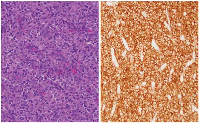
Histologic examination demonstrated a diffusely infiltrating glial neoplasm with high cellularity, marked nuclear pleomorphism, frequent mitotic figures including atypical forms, microvascular proliferation, focal pseudopalisading necrosis, and extensive geographic necrosis. Tumor cells showed diffuse positivity for GFAP and SOX2, supporting glial differentiation. ATRX expression was retained. The Ki-67 proliferation index reached up to 30 to 40 percent. IDH1 R132H and pan-TRK immunostains were negative. ALK (clone D5F3) immunohistochemistry demonstrated diffuse strong cytoplasmic positivity (Fig. 1). Notably, this staining pattern differs from that reported in epithelioid fibrous histiocytoma harboring PRKAR2A::ALK fusion, in which ALK expression characteristically shows a dot-like cytoplasmic pattern,3 whereas our case exhibited diffuse cytoplasmic staining.
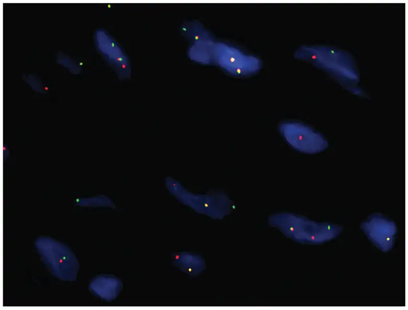
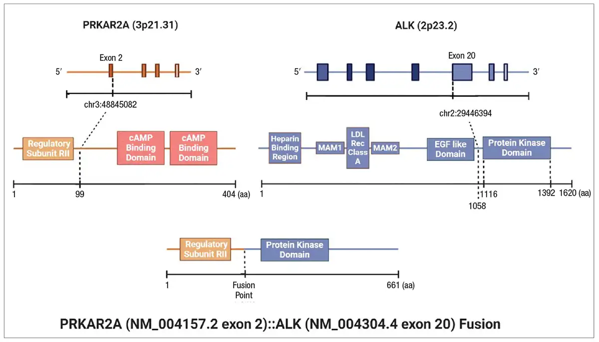
Given the diffuse ALK immunoreactivity, fluorescence in situ hybridization using an ALK (2p23) break-apart probe was performed and demonstrated rearrangement in 84.5 percent of analyzed tumor nuclei (Fig. 2). To further characterize the rearrangement, RNA-based next-generation sequencing was performed using an anchored multiplex PCR fusion panel. This analysis identified a predicted in-frame PRKAR2A (exon 2)::ALK (exon 20) fusion (Fig. 3).
Based on the histologic features and identification of an RTK fusion, the tumor was classified as infant-type hemispheric glioma. The patient received carboplatin and etoposide chemotherapy; however, interval imaging demonstrated tumor progression. Repeat surgical debulking was performed, and additional molecular studies were undertaken on the recurrent tumor.
Comprehensive DNA-based NGS using a 586-gene panel again identified the PRKAR2A::ALK fusion, with no additional pathogenic single nucleotide variants, insertions or deletions, copy number alterations, or structural rearrangements detected. Tumor mutation burden was low (1.14 mutations/Mb), and the tumor was microsatellite stable. Whole transcriptome sequencing confirmed expression of the fusion transcript and demonstrated aberrant activation of multiple signaling proteins, including TYK2, TNK2, EGFL7, MAPK11, and MAPK12. These genes have been implicated in immune regulation, tumor progression, angiogenesis, and oncogenic signaling pathways across a variety of malignancies.4,5 In particular, MAPK11 and MAPK12 belong to the p38 MAPK family, a component of the broader MAPK signaling network that is frequently dysregulated in cancer and has emerged as a therapeutic target in several neoplasms.6 While the clinical significance of these transcriptional alterations in infant-type hemispheric glioma remains uncertain, their activation raises the possibility of downstream MAPK pathway involvement and suggests potential avenues for future investigation into targeted therapies and immunomodulatory approaches. Overall, these findings further support the role of the PRKAR2A::ALK fusion as the primary oncogenic driver in this tumor.
Discussion. IHG is defined by the presence of RTK fusions that serve as dominant oncogenic events in an otherwise genomically stable tumor. Among these, ALK rearrangements represent a well-established subset; however, the diversity of ALK fusion partners in IHG continues to expand. The PRKAR2A::ALK fusion is a rare ALK rearrangement previously described in epithelioid fibrous histiocytoma,7 but, to our knowledge, it has not been reported in infant-type hemispheric glioma.
The fusion involves exons 1–2 of PRKAR2A, encoding a regulatory subunit of protein kinase A, fused in-frame to exon 20 of ALK, which encodes the tyrosine kinase domain. Although the functional consequences of this specific fusion have not been experimentally characterized, its structure is analogous to other activating ALK fusions and is predicted to result in constitutive kinase activation.8 Detection of the same fusion in both the primary and recurrent tumor, in the absence of additional pathogenic alterations, supports its role as an early driver event.
This case highlights the value of a multimodal molecular diagnostic approach. Diffuse ALK immunoreactivity served as an initial screening tool, while FISH confirmed gene rearrangement and the RNA-based NGS precisely identified the fusion partner and breakpoint. Subsequent DNA-based and transcriptome-level analyses provided orthogonal confirmation and demonstrated low genomic complexity, a hallmark of IHG. Such layered testing is particularly important in pediatric CNS tumors, where accurate molecular classification directly informs diagnosis and therapeutic consideration.
From a therapeutic standpoint, identification of ALK fusions raises the possibility of targeted kinase inhibition. However, clinical experience with ALK inhibitors in infants remains limited, and age-related regulatory constraints may delay treatment initiation. In IHGs, conventional chemotherapy appears largely ineffective with frequent recurrence reported.9-11 Therefore, consideration of ALK inhibitor therapy prior to one year of age may be warranted to improve outcomes.
In this case, the patient was started on single-agent oral lorlatinib (an ALK inhibitor) (25 mg daily)12 at age one year and has been doing well since initiation. Nevertheless, molecular identification of actionable fusions remains essential for guiding clinical trial enrollment and informing future therapeutic decision-making.
In conclusion, this case of IHG with a novel ALK fusion partner expands the known molecular spectrum of the disease. Transcriptome analysis demonstrated aberrant activation of five signaling proteins involved in cellular communication, four of which play key roles in intracellular signal transduction. From a management perspective, this case highlights the importance of considering early ALK inhibitor therapy given the limited efficacy of conventional chemotherapy. It also underscores the need for registry data and prospective studies to better define clinical outcomes and optimize management strategies in infants.
- Gianno F, Giovannoni I, Cafferata B, et al. Paediatric-type diffuse high-grade gliomas in the 5th CNS WHO Classification. Pathologica. 2022;114(6):422–435.
- Guerreiro Stucklin AS, Ryall S, Fukuoka K, et al. Alterations in ALK/ROS1/NTRK/MET drive a group of infantile hemispheric gliomas. Nat Commun. 2019;10(1):4343. doi.org/10.1038/s41467-019-12187-5
- Dawson K, Bridge JA, Sumegi J, Royce T, Gardner JM, Shalin SC. Epithelioid fibrous histiocytoma with dot-like perinuclear ALK expression and PRKAR2A-ALK fusion. Am J Dermatopathol. 2020;42(11):861–864.
- Sridaran D, Mahajan NP. ACK1/TNK2 kinase: molecular mechanisms and emerging cancer therapeutics. Trends Pharmacol Sci. 2025;46(1):62–77.
- Park S, Kim TH, Kim SH, You S, Jung Y. Three-dimensional vascularized lung cancer-on-a-chip with lung extracellular matrix hydrogels for in vitro screening. Cancers (Basel). 2021;13(16):3930. doi.org/10.3390/cancers13163930
- Phan T, Zhang XH, Rosen S, Melstrom LG. P38 kinase in gastrointestinal cancers. Cancer Gene Ther. 2023;30(9):1181–1189.
- Kazakov DV, Kyrpychova L, Martinek P, et al. ALK gene fusions in epithelioid fibrous histiocytoma: a study of 14 cases, with new histopathological findings. Am J Dermatopathol. 2018;40(11):805–814.
- Elshatlawy M, Sampson J, Clarke K, Bayliss R. EML4-ALK biology and drug resistance in non-small cell lung cancer: a new phase of discoveries. Mol Oncol. 2023;17(6):950–963.
- Shahab SW, Schniederjan M, Vega JV, et al. Case report: ATIC-ALK fusion in infant-type hemispheric glioma and response to lorlatinib. Front Oncol. 2023;13:1123378. doi.org/10.3389/fonc.2023.1123378
- Arthur C, Georgantzi K, de Ståhl TD, et al. Novel activating SNRNP70-ALK fusion in congenital infant-type hemispheric glioma displays clinical response to lorlatinib: a case-report. NPJ Precis Oncol. 2026;10:113. doi.org/10.1038/s41698-026-01336-x
- Greenwell AM, Baughan S, Altinok D, et al. Lorlatinib for the treatment of ALK fusion-positive infant-type hemispheric glioma: a case report. JCO Precis Oncol. 2022;6:e2200255. doi.org/10.1200/PO.22.00255
- Goldsmith KC, Park JR, Kayser K, et al. Lorlatinib with or without chemotherapy in ALK-driven refractory/relapsed neuroblastoma: phase 1 trial results. Nat Med. 2023;29:1092–1102.
Dr. Yilmaz and Dr. Zaheer were fellows in molecular genetic pathology at the time of the writing of this report; Dr. Soderquist is associate professor of pathology and cell biology; Dr. Mansukhani is professor of pathology and cell biology; and Dr. Hsiao is associate professor and Dr. Smithgall is assistant professor of pathology and cell biology—all in the Department of Pathology and Cell Biology, Columbia University College of Physicians and Surgeons. Dr. Yilmaz is now assistant professor of pathology, Icahn School of Medicine at Mount Sinai. Dr. Zaheer is assistant professor in the hematopathology and molecular genetic pathology divisions, University of Arkansas for Medical Sciences.