


Jonas J. Heymann, MD
Anupama Sharma, MD
Deepu Alex, MD, PhD
August 2026—Although cytologists and cytopathologists expend considerable effort in screening, risk stratification, diagnosis, staging, and prognostication, clinicians are increasingly demanding ancillary testing that predicts response or resistance to the broad range of therapeutic agents available to them. In advanced-stage non-small cell lung carcinoma, these therapies include tyrosine kinase and other small-molecule inhibitors, bispecific antibodies, and antibody-drug conjugates. They also include immune checkpoint inhibitors (ICI), which fall under the broader heading of immunotherapy. Here, we briefly review one such checkpoint—programmed death-1 (PD-1)—and one of its principal ligands, PD-L1.
Immune checkpoints are regulatory mechanisms that either stimulate or inhibit immune responses as the system attempts to distinguish self from non-self. PD-1 is an inhibitory checkpoint molecule expressed on both effector (Teff) and regulatory (Treg) T lymphocytes and, to a lesser extent, B lymphocytes, natural killer (NK) cells, monocytes, and Langerhans cells. PD-L1 is expressed on a broader subset of hematopoietic cells—including macrophages—and non-hematopoietic cells, including epithelial cells. PD-L1 expression may be induced in peripheral tissues or immune-privileged sites by proinflammatory cytokines, including type one and two interferons, tumor necrosis factor-α, and vascular endothelial growth factor. PD-L1 may also be constitutively expressed by tumor cells as a mechanism of immune escape: Its interaction with PD-1 acts as a “brake” on T lymphocytes within the tumor microenvironment, promoting Treg proliferation, converting Teff to Treg, and reducing global T lymphocyte production of interferon-γ, among other immunosuppressive effects.
ICIs derive their therapeutic benefit by disrupting the interaction between inhibitory checkpoint molecules and their ligands, thereby removing this “brake” and permitting anti-tumor Teff activity. Because Teff may attack normal tissues as well as tumor cells, ICI therapy carries autoimmune-like adverse events including dermatitis, enteritis, pneumonitis, hepatitis, and endocrinopathies. Multiple approved monoclonal antibody ICIs target either PD-1 (pembrolizumab, nivolumab, cemiplimab, dostarlimab, retifanlimab, toripalimab, tislelizumab) or PD-L1 (atezolizumab, durvalumab, avelumab, cosibelimab). PD-1 inhibitors additionally disrupt the interaction between PD-1 and PD-L2, expressed on antigen-presenting cells, though the clinical implications of this second ligand interaction remain less well understood. To avoid both unnecessary toxicity and the risk of withholding a more beneficial therapy, it is essential to identify patients whose tumors are most likely to respond to ICI.
Along with tumor mutation burden quantified by next-generation sequencing, PD-L1 expression by immunohistochemistry is FDA approved for predicting patient response to PD-1 or PD-L1 inhibitors. Currently unapproved approaches—such as transcriptional profiling and analysis of tumor-infiltrating lymphocytes—are emerging. PD-L1 IHC rests on the principle that tumors with broader constitutive PD-L1 expression are more likely to respond to ICI therapy, a hypothesis borne out by clinical trials despite some idiosyncratic responses. Similarly, tumors in which tumor-infiltrating lymphocytes and other immune cells express PD-L1 are also associated with higher response rates. Unfortunately, the methods by which PD-L1 is quantified are far from uniform.
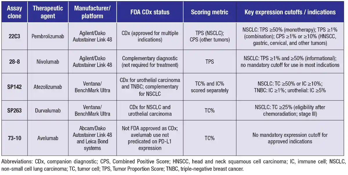
Antibodies available for quantification of PD-L1. The pharmaceutical companies that developed therapeutic PD-1 and PD-L1 monoclonal antibodies also developed predictive IHC assays for each respective therapeutic agent in tandem. Although all assays quantify PD-L1 expression by IHC, each uses its own monoclonal antibody clone, immunostaining platform, scoring system, protocol, and expression-level cutoffs corresponding to specific therapeutic indications (Table 1). Some assays received FDA companion diagnostic (CDx) designation; others did not, or received CDx approval only for certain indications. The potential lack of interchangeability among assays presents a figurative Gordian knot for pathologists.
The industry-supported Blueprint PD-L1 IHC Assay Comparability (“Blueprint”) Project, which reported its results in two phases, attempted to gently untie the proverbial knot. In phase one, four assays (22C3, 28-8, SP142, SP263) were directly compared on serial sections of 39 NSCLC resection specimens.1 Although a different classification would have been assigned in 37 percent of cases depending on which assay was used, the percentage of PD-L1 staining in tumor cells was comparable among three of the four assays. SP142 was less sensitive for PD-L1 detection and demonstrated weaker staining intensity. All four assays showed inconsistency when evaluating immune cell staining. A concurrent harmonization study demonstrated similar intraobserver variability among assays for both tumor and immune cells.2
Phase two of the Blueprint Project validated these findings using 71 clinical NSCLC samples—including 20 cytologic cell blocks—evaluated by 24 pathologists from 15 countries across five continents and expanded the analysis to include the 73-10 assay and metastatic tumors.3 Tumor cell PD-L1 staining remained comparable among three assays. SP142 was again less sensitive than 22C3, 28-8, and SP263, while 73-10 was found to be more sensitive. Immune cell staining remained inconsistent across all assays. Critically, interobserver agreement for tumor cell staining in cytologic samples was only slightly lower than for histologic samples, reaching at least moderate agreement (κ > 0.6) for 22C3, 28-8, and SP263 at all cutoffs. Together, the Blueprint Project results suggest that laboratories may use any one of the developed assays, provided they perform full validation as they would for any laboratory-developed test. The CAP offers proficiency testing programs for PD-L1 IHC to support this process.
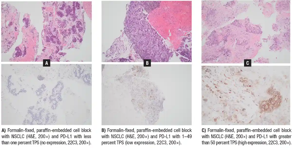
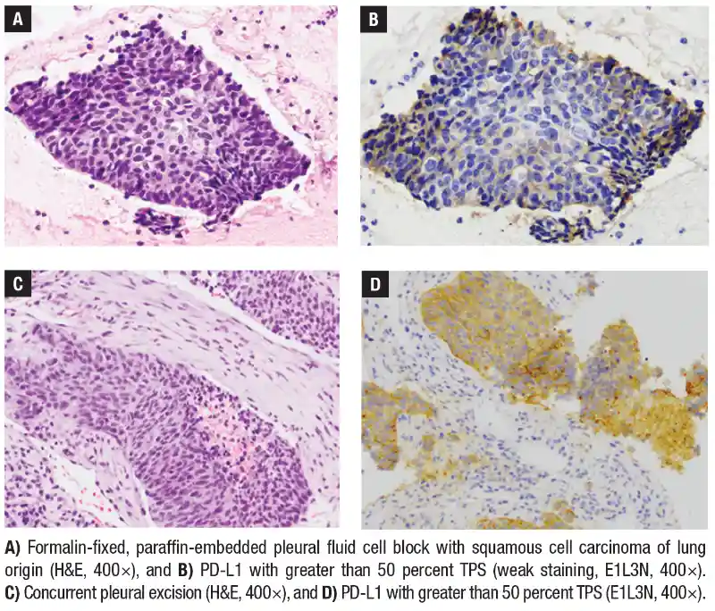
Numerous studies since the Blueprint Project have compared PD-L1 expression across resection specimens, cytologic cell blocks, and small biopsy specimens—comparisons that are critical because cytology and small biopsy specimens often represent the only available tissue in patients with advanced, unresectable disease. Results have been widely variable. One early study using SP142 and E1L3N in NSCLC tissue sections offered a partial explanation: McLaughlin, et al., observed positive staining in some tumor regions but negative staining in others, documenting spatial heterogeneity.4 Subsequent studies confirmed spatial heterogeneity and further revealed significant temporal heterogeneity in PD-L1 expression. Despite this variability, the best available evidence—including a meta-analysis encompassing non-cell block cytologic specimens and small histologic biopsies such as core needle biopsies—indicates that IHC on cytologic specimens provides accurate PD-L1 assessment compared with histologic specimens for NSCLC at both ≥ one percent and ≥50 percent cutoffs.5 Some studies suggest that cytologic cell blocks may even outperform small histologic biopsies for this purpose,6 though neither matches the accuracy of resection specimens.
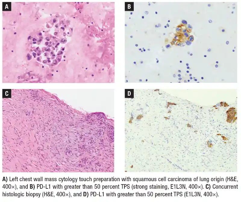
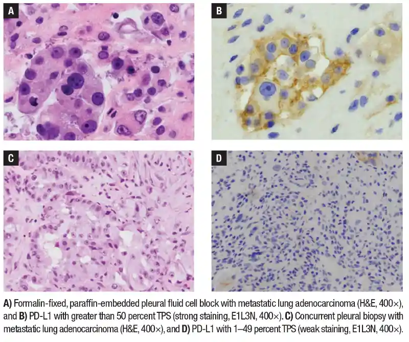
Interpretation of PD-L1 IHC. To identify NSCLC tumors likely to respond to PD-1 or PD-L1 inhibitors, pathologists calculate the Tumor Proportion Score (TPS): the percentage of all viable tumor cells exhibiting partial or complete membranous PD-L1 staining. TPS equals the number of staining tumor cells divided by the total number of viable tumor cells. A minimum of 100 tumor cells is required, though 200 or more is advisable when TPS falls near a clinical cutoff (one percent or 50 percent). Cytologic cell blocks are well suited to TPS calculation because NSCLC tumor cells are generally easy to identify. A critical point is the exclusion of immune cells—particularly macrophages, which characteristically show strong and diffuse PD-L1 expression.
For tumor types other than NSCLC, pathologists calculate the Combined Positive Score (CPS): the ratio of all PD-L1-expressing cells—including tumor cells and tumor-associated immune cells—to the total number of viable tumor cells, multiplied by 100. CPS is more challenging to apply to cytologic cell blocks because the disaggregation of tumor cells from stroma makes it difficult to determine which immune cells are truly tumor associated, a problem compounded in immune-rich sites such as lymph nodes. Nevertheless, limited evidence supports CPS calculation on cytologic cell blocks in selected circumstances.7
Future directions: immune checkpoints beyond PD-1. CTLA-4, LAG-3, TIM-3, and TIGIT are inhibitory immune checkpoints for which pharmacologic inhibitors are either currently FDA approved or under active development for NSCLC and other malignancies. Ipilimumab, a CTLA-4 inhibitor whose availability predates that of PD-1 and PD-L1 inhibitors, holds numerous therapeutic indications. For metastatic or recurrent NSCLC lacking a targetable genetic alteration, ipilimumab is FDA approved in combination with nivolumab, with or without platinum-based chemotherapy, depending on PD-L1 expression.8,9 Tremelimumab, a newer CTLA-4 inhibitor, is FDA approved for metastatic NSCLC in combination with durvalumab and platinum-based chemotherapy; its use is predicated on neither CTLA-4 expression nor expression of its ligands, CD80 and CD86. Relatlimab, a LAG-3 inhibitor, carries an FDA-approved indication for metastatic melanoma irrespective of LAG-3 expression. No TIM-3 or TIGIT inhibitors have yet received FDA approval, though both classes remain under investigation and may expand the landscape of immune checkpoint therapy in the future.
- Hirsch FR, McElhinny A, Stanforth D, et al. PD-L1 immunohistochemistry assays for lung cancer: results from phase 1 of the Blueprint PD-L1 IHC Assay Comparison Project. J Thorac Oncol. 2017;12(2):208–222.
- Scheel AH, Dietel M, Heukamp LC, et al. Harmonized PD-L1 immunohistochemistry for pulmonary squamous-cell and adenocarcinomas. Mod Pathol. 2016;29(10):1165–1172.
- Tsao MS, Kerr KM, Kockx M, et al. PD-L1 immunohistochemistry comparability study in real-life clinical samples: results of Blueprint Phase 2 Project. J Thorac Oncol. 2018;13(9):1302–1311.
- McLaughlin J, Han G, Schalper KA, et al. Quantitative assessment of the heterogeneity of PD-L1 expression in non-small-cell lung cancer. JAMA Oncol. 2016;2(1):46–54.
- Tajarernmuang P, Aliaga F, Alwakeel AJ, et al. Accuracy of cytologic vs histologic specimens for assessment of programmed cell death ligand-1 expression in non-small cell lung cancer: a systematic review and meta-analysis. Chest. 2024;165(2):461–474.
- Heymann JJ, Bulman WA, Swinarski D, et al. PD-L1 expression in non-small cell lung carcinoma: comparison among cytology, small biopsy, and surgical resection specimens. Cancer Cytopathol. 2017;125(12):896–907.
- Liu Z, Williams M, Stewart J, Glisson BS, Fuller C, Roy-Chowdhuri S. Evaluation of programmed death ligand 1 expression in cytology to determine eligibility for immune checkpoint inhibitor therapy in patients with head and neck squamous cell carcinoma. Cancer Cytopathol. 2022;130(2):110–119.
- Paz-Ares LG, Ramalingam SS, Ciuleanu TE, et al. First-line nivolumab plus ipilimumab in advanced NSCLC: 4-year outcomes from the randomized, open-label, phase 3 CheckMate 227 Part 1 trial. J Thorac Oncol. 2022;17(2):289–308.
- Paz-Ares L, Ciuleanu TE, Cobo M, et al. First-line nivolumab plus ipilimumab combined with two cycles of chemotherapy in patients with non-small-cell lung cancer (CheckMate 9LA): an international, randomised, open-label, phase 3 trial. Lancet Oncol. 2021;22(2):198–211.
Dr. Heymann is an associate professor of clinical pathology and laboratory medicine at Weill Cornell Medicine, New York, NY. Dr. Sharma is a staff pathologist at Sentara Health, Norfolk, Va. Dr. Alex is a clinical associate professor of pathology and laboratory medicine, University of British Columbia, and a member of the British Columbia Cancer Agency, Vancouver, BC. All are members of the CAP Cytopathology Committee.