


M. Lisa Zhang, MD
Ivan Chebib, MD
August 2026—Cytology specimens have long played a central role in primary diagnosis, with fine-needle aspiration often serving as the first—and sometimes only—procedure performed before treatment decisions are made. However, when molecular testing is needed, cytology material is still often viewed through a histology-centered lens: Is there a cell block or core biopsy? This emphasis can overlook one of the most cellular and diagnostically informative components of the specimen: the direct smear.
The utility of cytology specimens for molecular testing is well established, particularly in lung cancer, where cytopathologists have become accustomed to triaging small samples for biomarker testing. As previously highlighted by Makarenko and Kim, cytology specimens can support a broad range of molecular assays when preanalytic variables, tumor cellularity, and laboratory validation are appropriately addressed.1 But the role of cytology smears extends beyond “getting enough material” for next-generation sequencing panels. In selected cases, smear slides can provide material for molecular tests that directly establish the primary diagnosis.
This distinction is important. Molecular testing in cytology is often discussed in the context of predictive biomarker testing, such as for EGFR, ALK, ROS1, RET, NTRK, and other therapeutically relevant alterations. However, many tumors, particularly salivary gland and soft tissue neoplasms, are now defined or refined by recurrent gene fusions. In these settings, molecular testing is not merely ancillary to a diagnosis already made by morphology; rather, it may be the only evidence that moves a case from an indeterminate diagnostic category to the diagnosis of a specific entity.
Previous work by Ramani, et al., helped establish that cytology smears can improve the success of RNA-based NGS fusion testing.2 In that study, the addition of direct smears substantially increased adequacy for RNA-based fusion testing from 45 percent (cell block alone) to 92 percent, supporting the concept that smear material can be a robust substrate when formalin-fixed, paraffin-embedded material is limited.
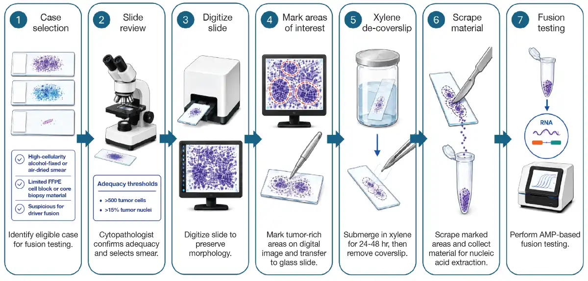
Wang, et al., recently demonstrated the diagnostic potential of sacrificed cytology smears for anchored multiplex PCR-based fusion testing.3 In that series, direct smears were used when FFPE cell block or core biopsy material was insufficient but highly cellular cytology smears were available. Before sacrifice, the selected slide was digitally scanned to preserve a permanent morphologic record. Tumor-rich areas were marked, coverslips were removed, and cellular material was scraped for RNA-based fusion testing (Fig. 1). Across 14 cases tested using scraped smears, adequate nucleic acid was obtained in all cases, and fusions were detected in nine cases (64 percent). Importantly, the results allowed for definitive tumor classification and primary diagnosis of the following entities: adenoid cystic carcinoma (MYB::NFIB), mucoepidermoid carcinoma (CRTC1::MAML2), pleomorphic adenoma (ACTB::PLAG1, NCALD::PLAG1), dermatofibrosarcoma protuberans (COL1A1::PDGFB), and nodular fasciitis (COL1A2::USP6).
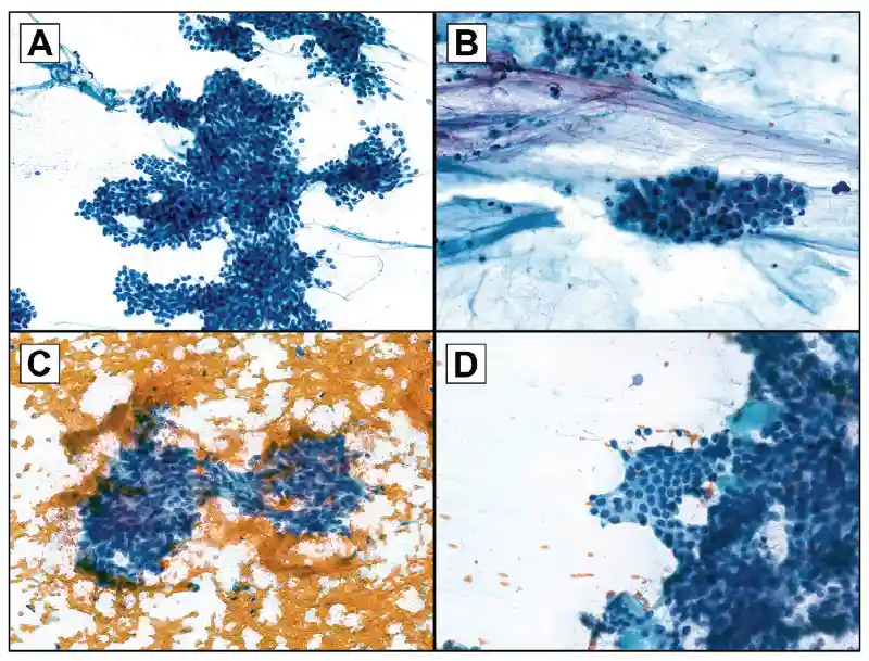
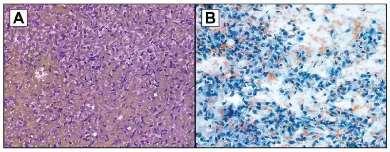
These examples underscore the value of cytology smears for informing patient management. A PLAG1 or MYB::NFIB fusion can make the important distinction between a benign pleomorphic adenoma and a malignant adenoid cystic carcinoma, respectively, when the cytology diagnosis would otherwise be “basaloid neoplasm” (Fig. 2). In the soft tissue realm, the identification of a sarcoma requiring surgical removal versus nodular fasciitis can potentially avoid overtreatment of a self-limited lesion (Fig. 3). These molecular results can change the diagnostic category, guide surgical planning, or support conservative management.
There are, of course, important caveats. A smear that is sacrificed for molecular testing is no longer available as a glass slide for future review, which is why digital archiving before scraping is critical. Laboratories must validate the specific assay and specimen type, establish minimum cellularity and tumor fraction thresholds, and define quality metrics for reporting, particularly for negative results. A negative fusion assay does not exclude a diagnosis if the tumor may harbor alternative fusions, noncanonical breakpoints, or other molecular mechanisms. Cytomorphology, imaging, clinical findings, and surgical pathology correlation remain essential.
As molecular testing becomes increasingly integrated into cytopathology practice, direct smears deserve renewed attention as a diagnostically valuable specimen type. When cell block material is limited, a well-prepared cellular smear may be the highest-yield specimen available. With appropriate validation, digital preservation, and careful case selection, cytology smears can help deliver not only predictive biomarker results but also definitive primary diagnoses.n
- Makarenko VV, Kim AS. Doing a lot with a little: molecular testing on cytology specimens. College of American Pathologists. June 28, 2022. https://www.cap.org/member-resources/articles/doing-a-lot-with-a-little-molecular-testing-on-cytology-specimens
- Ramani NS, Chen H, Broaddus RR, et al. Utilization of cytology smears improves success rates of RNA-based next-generation sequencing gene fusion assays for clinically relevant predictive biomarkers. Cancer Cytopathol. 2021;129(5):374–382.
- Wang CI, Zhang ML, Madrigal E, Faquin WC, Chebib I. Diagnostic utility of anchored multiplex polymerase chain reaction-based fusion assay on sacrificed cytology slides. Cancer Cytopathol. 2026;134(6):e70105.
Dr. Zhang is a cytopathologist and gastrointestinal pathologist, Massachusetts General Hospital, and assistant professor of pathology, Harvard Medical School. Dr. Zhang is a member of the CAP Cytopathology Committee. Dr. Chebib is a cytopathologist and soft tissue pathologist, Massachusetts General Hospital, and associate professor of pathology, Harvard Medical School.