


Lamé Balikani, MD, MPH
August 2026—Pediatric pathology remains a relatively small subspecialty within anatomic pathology. The American Board of Pathology since 1990 has issued only 648 subspecialty certificates in pediatric pathology, and between 2020 and 2024, only 14 to 21 candidates sat for the pediatric pathology certifying examination each year. Furthermore, even fewer pathologists hold subspecialty certification in both pediatric pathology and cytopathology. Interestingly, during the May 2024 review of active certifications, ABPath identified only 31 pathologists nationwide with active certifications in both subspecialties.
Despite the limited number of these dual-certified pathologists, pediatric cytopathology services are routinely staffed by pediatric pathologists with specialized expertise in cytology, cytopathologists practicing in pediatric-focused institutions, and cytopathologists in mixed adult-pediatric practice settings who regularly evaluate specimens from children. As a result, these overlapping topics are relevant to many practicing pathologists.
Most cytopathology training programs are based in hospitals treating adults, with few programs emphasizing both adult and pediatric laboratory samples. Because exposure to pediatric cytology is limited, it raises the question: What distinguishes pediatric cytology (especially in exclusive pediatric contexts)?
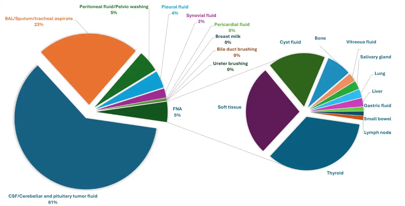
1. Relatively fewer cases. Data from a single institution reports 1,500 to 1,700 annual cases, with little to no gynecologic cytology samples. Most specimens are exfoliative. Of these, cerebrospinal fluid (CSF) is the most common type, followed by bronchoalveolar lavage and peritoneal/pleural/pericardial fluid. Fine-needle aspiration (FNA) specimens make up about five percent of the total (Fig. 1).
2. Fine-needle aspiration. In children, FNAs are mostly performed in interventional radiology (IR). These are done under anesthesia or sedation. Some adolescent patients undergo FNA under local anesthesia in IR, at the bedside, or in the ear, nose, and throat clinic.
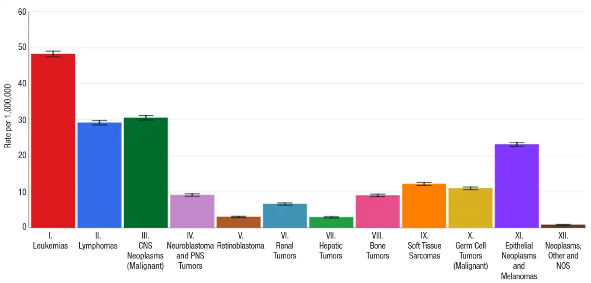
In pediatric patients, diagnostic considerations and differential diagnoses diverge from those in adults, requiring pathologists to be vigilant. The most common malignancies include hematolymphoid and central nervous system tumors followed by epithelial neoplasms among others (Fig. 2). Furthermore, the incidence and type of malignancy vary with age.
In thyroid cytology, there are several important differences in pediatric patients compared with adults:
- Thyroid nodules are more common in adults (four percent–67 percent) compared with children (one percent–13 percent).
- The risk of malignancy in pediatric thyroid nodules (22 percent–26 percent) is higher than in adult nodules (five percent–15 percent). This increased risk is especially pronounced in the indeterminate categories of the Bethesda System for Reporting Thyroid Cytopathology.
- Children with papillary thyroid carcinoma (PTC) are more likely to have regional lymph node involvement, extrathyroidal extension, and pulmonary metastasis.
- PTC is the most common pediatric thyroid carcinoma, as in adults. In children, the tumor is more often associated with molecular fusion (such as RET and NTRK fusions). Relatively fewer cases have BRAF mutations, which are more common in adults.
- Syndromes are important in pediatric pathology. Relevant syndromes in thyroid pathology include MEN 2A/2B/Familial MTC, Carney complex, DICER1, Familial Adenomatous Polyposis, Cowden/PTEN hamartoma, and McCune-Albright. DICER1 syndrome in the thyroid is associated with early-onset multinodular goiter, follicular-patterned tumors (adenomas and carcinomas). Pediatric papillary thyroid carcinomas may harbor somatic DICER1 mutations. While thyroid carcinomas with underlying DICER1 mutations are usually indolent, recent studies have shown that pediatric poorly differentiated thyroid carcinoma and thyroblastoma can also be seen.
3. Interesting cytology cases.
- CSF: In addition to CSF sampling for possible infections, CSF evaluation is required for the staging and monitoring of treatment response in patients with leukemias and CNS tumors. Although CSF is the most common specimen type in some pediatric cytology practices, it is rarely positive for malignancy (less than two percent).
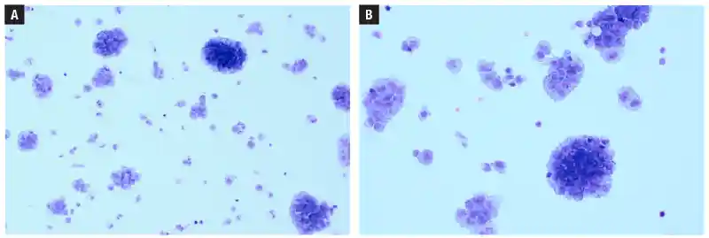
Fig. 3 shows cytospin from a CSF sample obtained from a 15-month-old female with a history of a cerebellar lesion with germline SMARCA4 gene duplication and an atypical teratoid/rhabdoid tumor (ATRT).
The Giemsa-stained cytospin shows a cellular sample composed of large plasmacytoid cells with abundant cytoplasm, eccentrically placed nuclei, and prominent nucleoli, consistent with the patient’s known history of ATRT.
- Pleural fluid. Fig. 4 shows pleural fluid obtained from a 12-month-old male with a history of a chest wall mass involving soft tissue and vascular structures, as well as prominent mediastinal lymphadenopathy.
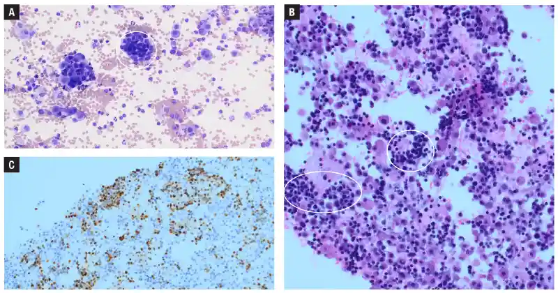
The Giemsa-stained cytospin is highly cellular and revealed variably sized three-dimensional clusters composed of atypical cells in a background of reactive mesothelial cells, histiocytes, and lymphocytes. At higher magnification, the atypical cells have a high nuclear to cytoplasmic (N/C) ratio, with irregular nuclear borders, and scant blue to amphophilic cytoplasm. The H&E-stained cell block shows clusters and single cells with a high N/C ratio, hyperchromatic nuclei, and scant eosinophilic cytoplasm in a background of reactive mesothelial cells, histiocytes, and lymphocytes. The atypical cells show diffuse nuclear positivity for myogenin (Fig. 4c), consistent with biopsy-proven rhabdomyosarcoma.
Rhabdomyosarcoma cells in fluid have variable morphologic features. They range from small round cells with scant cytoplasm to cells with more abundant cytoplasm, imparting rhabdoid appearance. In some cases, the rhabdomyosarcoma cells resemble mesothelial cells or histiocytes. For small round cell tumors—such as lymphoma, neuroblastoma, or Wilms tumor—clinical history and immunohistochemical stains help establish the correct diagnosis.
- Primary screening. Due to low specimen counts, some pediatric-only practices—especially those without gynecologic cytology—have few cytologists for screening. Pathologists may need to serve as primary screeners. This practice complicates workflow and may require discussing challenging cases with colleagues or double-reviewing certain cases after a “washout” period.
In summary, pediatric cytology is a challenging and distinct area of practice. Differences in specimen types, disease prevalence, molecular alterations, hereditary syndromes, and laboratory workflow distinguish pediatric-based specimens from those encountered in most pathologists’ routine practice. As a result, cytopathologists should recognize the distinct features of pediatric cytology and understand when ancillary studies, multidisciplinary discussion, and subspecialty consultation can improve patient care.n
- American Board of Pathology. 2024 annual report. Published June 23, 2025. www.abpath.org/wp-content/uploads/2025/06/ABPath-2024-Annual-Report_final.pdf
- American Board of Pathology. Board certifications in pediatric pathology and cytopathology. Personal communication.
- National Cancer Institute. National Childhood Cancer Registry Explorer. https://nccrexplorer.ccdi.cancer.gov
- Siegel RL, Miller KD, Fuchs HE, Jemal A. Cancer statistics, 2021. CA Cancer J Clin. 2021;71(1):7–33.
- Francis GL, Waguespack SG, Bauer AJ, et al. Management guidelines for children with thyroid nodules and differentiated thyroid cancer: the American Thyroid Association Guidelines Task Force on pediatric thyroid cancer. Thyroid. 2015;25(7):716–759.
- Gupta A, Ly S, Castroneves LA, et al. A standardized assessment of thyroid nodules in children confirms higher cancer prevalence than in adults. J Clin Endocrinol Metab. 2013;98(8):3238–3245.
- Paulson VA, Rudzinski ER, Hawkins DS. Thyroid cancer in the pediatric population. Genes (Basel). 2019;10(9):723. doi.org/10.3390/genes10090723
- Ali SZ, VanderLaan PA, eds. The Bethesda System for Reporting Thyroid Cytopathology: Definitions, Criteria, and Explanatory Notes. 3rd ed. Springer; 2023.
- Sauer M, Barletta JA. Proceedings of the North American Society of Head and Neck Pathology, Los Angeles, CA, March 20, 2022: DICER1-related thyroid tumors. Head Neck Pathol. 2022;16(1):190–199.
- Fuller MY, Shrestha S, Baskota SU. International perspective on pediatric cytology. Cancer Cytopathol. 2025;133(1):e22911.
- Saoud C, Ali SZ. Metastatic sarcomas to pleural effusion: a 10-year large tertiary care center experience with emphasis on clinical features and cytomorphologic characteristics. J Am Soc Cytopathol. 2023;12(3):216–228.
Dr. Balikani is a staff pathologist, Nationwide Children’s Hospital, Columbus, Ohio. She is a member of the CAP Cytopathology Committee.