


Amy Carpenter
October 2026—Clinical laboratory scientists in Cleveland Clinic’s parasitology laboratory were several months into implementing an AI-driven digital platform for ova and parasite testing when a historic cyclosporiasis outbreak swept through Ohio and other states this summer. When daily cases spiked into the hundreds in mid-July, they scaled up their Cyclospora cayetanensis testing capacity, powered by Techcyte’s AI-assisted screening tool.
“This is truly the fairytale of AI,” said Anisha Misra, PhD, director of parasitology and mycology at Cleveland Clinic and assistant professor of pathology, Cleveland Clinic Ler-ner College of Medicine. By the time Dr. Misra spoke in an ADLM annual meeting session in late July, her laboratory had tested 3,730 samples, of which 39 percent were positive. “Just by being able to implement AI,” she said.
Though scaling up manual microscopy is not easy, she and her colleagues were able to do it, thanks to artificial intelligence and “all the lessons learned over the last two years of trying to get it through the door,” she said.
The Techcyte algorithm, which is intended for trichrome smears, had already gone through a training validation when it came to Cleveland Clinic as a locked algorithm system, Dr. Misra said. She and her colleagues validated Blastocystis sp., Giardia duodenalis, Dientamoeba fragilis, Endolimax nana, Iodamoeba bütschlii, Entamoeba hartmanni, and Entamoeba sp. They chose not to validate C. cayetanensis (more on that later) and white and red blood cells.
They purchased the Hamamatsu NanoZoomer S360 scanner for the validation and the Sakura Finetek Tissue-Tek automated coverslipper.
They made preanalytic changes, one of which was to switch from a low-viscosity polyvinyl alcohol to a single-vial fixative to smooth out their processes. “We also changed our concentration steps,” she said, from manual to a Parasep vial.
One of the biggest challenges in implementing AI was standardizing how the laboratory created its slides. “We went from making hills and valleys for our trichrome stain to trying to get an even monolayer.” That’s important and dependent on the scanner, she said: “Our Hamamatsu scanner does not do the Z-stacking you would like. Pairing your workflows with the equipment you have becomes really important.”
Next up was training. “AI does not tell you whether there’s an organism present,” Dr. Misra said. “It puts up images and says, ‘I think there are this many Giardias present,’ it shows you those images, and it posts them at a confidence level with the best images on top and the lowest-confidence images on the bottom.”
The user must then discriminate whether the images are real by going into the algorithm and agreeing or disagreeing with the AI calls. “So even the AI call is with the human in the loop and the human is deciding,” she said.
How would the humans at Cleveland Clinic interact with the AI? To find out, Dr. Misra and colleagues curated a set of 154 positive and negative slides, aiming for as close to a 50/50 split as possible with all of the organisms represented within the data set. A single scan was generated and three parasitologists read it. “Same scan, no changes, same monolayer.”
If the scan was too thin or an incorrect area was scanned, they would call it invalid and push it toward a manual review.
Of the 154 slides, the total slide numbers the three parasitologists reviewed manually were 70, 82, and 101. The upshot of the exercise: The three parasitologists had different review behaviors with the same slides and same AI output. And nearly half of all negative slides were reviewed in one instance, which negates the intended efficiency of AI.
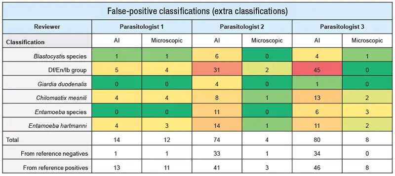
The team also looked for false-positive classifications (classified as extra classifications) (Fig. 1). Some organisms, they found, were more prone to being called by AI. “AI makes gray zones grayer,” she said. Giardia and Blastocystis, with their concrete shape, are detectable. The amorphous organisms, like Dientamoeba fragilis, are not, she said, “and that influences the human read. That’s where we saw the most amount of variation.” A manual review corrected most of those.
She pointed out that AI did not pick up positives in completely negative slides. “It picked up extra organisms in slides that were already positive for other organisms. We’re not sure what to make of that, but it’s something we noted in our data.”
Were the extra classifications truly extra classifications? “What if the human eye was missing the data?” Dr. Misra asked. There is variation between the AI and human reads, she said. AI can be a little more sensitive than a human, “and it does depend on how long the parasitologist has been training on the bench and how much experience they have.”
When the parasitologists went back to read the extra classification slides without knowing what the AI read was, all extra classifications went away. “That tells us the reader was influenced by what they were seeing on the AI read and then picking up on it.”
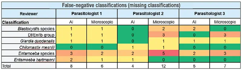
They looked, too, at missing (false-negative) classifications (Fig. 2) because the aim was to move to an algorithm that could rule out negative slides. “In our parasitology lab, our percent positivity is around two to maybe three percent in the best of years,” though not this year owing only to Cyclospora. “Because of that, we don’t want to spend the time reading negatives.”
Missed classifications were found to be infrequent, and when it was missed it was because the human reviewing the AI classified it as junk rather than a true organism. “So we noted that and said this is a risk with implementing AI and we’ll have to come up with mitigation strategies to move forward.”
Quality monitoring and clinical validation were the next step. All of the ova and parasites for six months were put through Techcyte to see how parasitologists would adjust to the workflow. “We had some cases reviewed simultaneously using both the AI platform and trichrome microscopy. In other cases, the AI-assisted and trichrome interpretations were performed independently so that we could evaluate the potential impact of reader bias.”
Most of the time, AI and the manual read were consistent for those six months, Dr. Misra said, “and what we had to do was apply consistent application of scan validity criteria,” meaning whenever errors were seen it was because the scan quality was poor. Degraded or suboptimal specimens were excluded from Techcyte review.
“Creating those monolayers has been the most challenging thing about implementing AI because if that scan is bad, then everything downstream is terrible.”
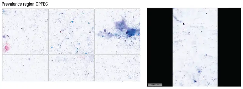
To reduce some of the variation in the parasitologists’ calls of invalid scans, they added criteria. “We look at our Hamamatsu scan, we make sure at least 10 of the 14 areas are zoomed in properly. We also looked at the prevalence region” (Fig. 3). The Techcyte algorithm divides a scan into 14 prevalence regions; if more than three are blurry, the laboratory called the scan invalid. “Those are the rules we’ve set in our lab to mitigate workflow risk.”
They manually confirm all positive results before reporting them. Cases with unusual, low count, or questionable morphology are escalated to the medical director for review.
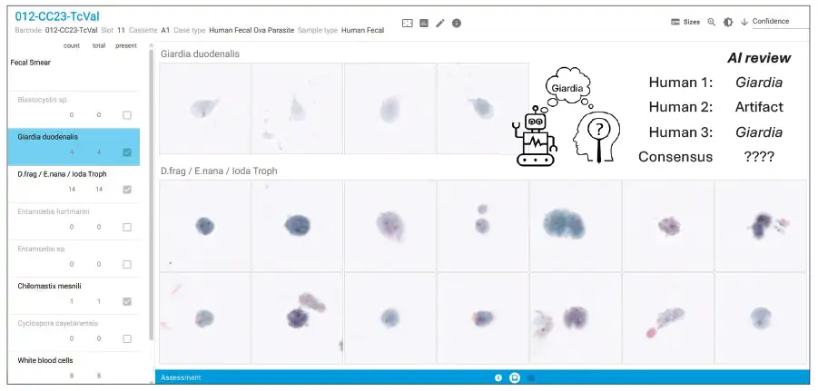
Fig. 4 (page 26) is an example; it’s a Techcyte read of a fecal smear trichrome stain in which the AI picked up four Giardia. When three humans read the stain, two called it positive for Giardia; one called it an artifact. “When they tried to do a manual review, they couldn’t find it the first time around,” Dr. Misra said.
What to do with the slides in question? “We’ve been setting them aside for the day and going back to review them,” she said, and until now, “we found all cases to be positive after extended review.” Thus, AI is increasing their sensitivity. “These are challenging and bring up the question of whether this is clinically relevant,” which she said is a separate talk for another time. “But this is our process”: review by the medical director or a more experienced parasitologist.
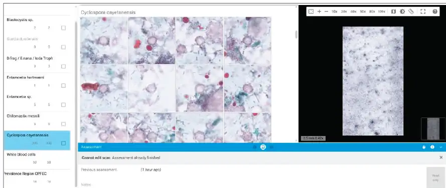
In early June this year, laboratory staff came across a case with C. cayetanensis (Fig. 5). The technologist reached out, concerned the laboratory hadn’t validated Cyclospora. “At the time we didn’t have enough specimens,” Dr. Misra explained. However, the laboratory had a workflow set up such that if C. cayetanensis was detected, medical director review would be needed, and upon discussion with the clinical team they would perform a modified acid-fast stain.
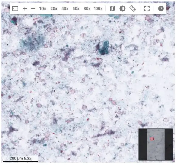
The modified acid-fast stain and autofluorescence were negative. “It was starch cells,” she said. In Fig. 6 are various sizes of starch cells that the AI platform identified as C. cayetanensis.
“This is where human judgment becomes extremely critical,” Dr. Misra said. “Even for the positives, even when they look great, you want to employ some degree of human assessment in order to report it out, especially when we’re not confident or have not validated that target.”
They also implemented a threshold-based reviewer review using minimum AI object count triggers and periodic secondary reviews of negative slides that met the threshold. “We found 398 Cyclospora. That’s what we’re calling our threshold of detection,” she said. The laboratory’s threshold is low for most organisms with defined shapes, like Giardia. Any organisms confidently detected by AI and confirmed by a human go through a secondary review. For amorphous organisms such as Dientamoeba, the threshold is higher, about 150. These data are pulled biweekly so they can assess which slides need a manual review that might not have been reviewed manually during their regular process. “This is the quality monitoring step that’s occurring in the background,” she said.
What exactly is a threshold, given it’s not a term used often in the context of AI algorithms and detections? “We wanted to set a baseline,” she said. To monitor the thresholds, they duplicated molecular testing. “We took a high-control slide with Blasto and a low-control slide with Giardia and we run it every day that we perform our OVAP trichrome staining. This allows us to monitor if our thresholds are staying consistent on the same slide, and as we change slides every three months because of photo-bleaching, we set thresholds again after 20 scans.”
Westgard rules are used to escalate cases to the medical director when concerning trends are seen.
When they saw a downward trend in Blastocystis, they investigated and found that the Hamamatsu scanner tends to adjust and learn where it needs to scan, and thus they figured out they needed to autocalibrate weekly using a blank slide. “That is unique to our scanner and something important for us to learn,” Dr. Misra said. Once they began to autocalibrate and run the same slides, they saw their control thresholds jump up. “We can monitor them. We ran the scans for 20 times and set a new threshold.”
The laboratory’s QA monitoring includes misclassification rates and object counts. “We take all the raw data we’re getting from our AI module and plot percent positivity, percent negativity, and our invalid rates to keep track of it,” Dr. Misra said, noting it’s the first time the laboratory has “this much nuance” in its monitoring. It’s important, she said, “because we saw when our percent positivity calls on AI decreased below that 10 percent threshold we would ideally like, we had three false-negatives when we reviewed the slides.”
Individual parasitologist metrics are also among those monitored. “As much as we’re looking at it as a group,” she said, “it was important to know where to step in and guide better use of the AI tool.” Newly hired parasitologists are trained with AI in mind and thus “their metrics are more balanced because they’re not coming in with previous behaviors.” Overall, she said, the use of AI has standardized reader training and competency assessment.
Turnaround time took a hit when AI was implemented. “All we did was add another step” for the parasitologists. So they studied and reevaluated the whole process and over time brought turnaround time to where it needed to be. “AI is just a tool, not a magical answer to improved turnaround times,” she said.
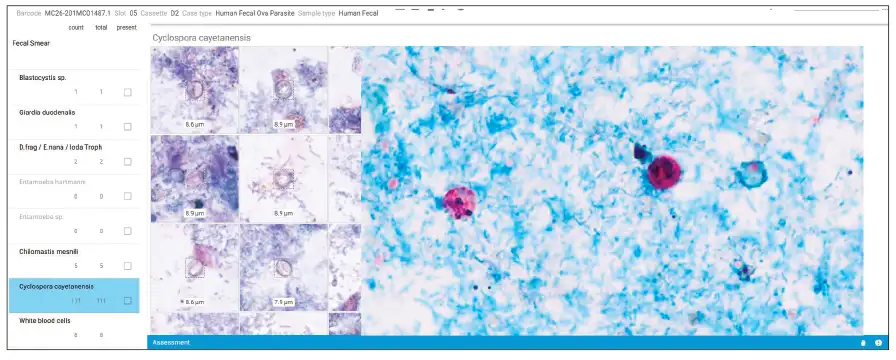
In late June, a parasitologist said she thought she was seeing a case of Cyclospora (Fig. 7). Dr. Misra replied: “We’ve been burned before. Let’s do the modified acid-fast stain; it’s probably starch cells.” The modified acid-fast stain was positive.
“During the next couple of days, we started getting more and more positives with our trichrome staining, and I was making more and more calls every day, and this piqued my curiosity. We were having conversations with emergency departments, family health centers, and our urgent care centers, and they were seeing more diarrhea cases, and we were seeing more Cyclospora.”
In the initial days of the outbreak, in late June, the laboratory was detecting all cases of C. cayetanensis using its trichrome stain. All such detections were followed by a call to the provider to say the AI algorithm was picking up Cyclospora but a confirmatory test was needed.
By July 1, a Cleveland Clinic notice warned of a possible outbreak and advised clinicians to order the modified acid-fast stain when seeing patients with gastrointestinal symptoms. “We had no idea at the time what we were in for,” Dr. Misra said.
Soon the laboratory was receiving 45 to 60 modified acid-fast stain samples a day and had to figure out how to scale up.
An algorithm was available, but C. cayetanensis detection on trichrome staining requires an additional modified acid-fast stain. “That seemed like a lot of work when we were trying to reduce work.” They therefore decided to validate the modified acid-fast stain for Cyclospora.
The AI is not trained to do that, Dr. Misra noted, but even on the trichrome stain, “the AI is picking up ghost cells,” not the faintly stained cells, “because of the cell wall of Cyclospora. So we thought it was a journey worth taking to figure out if it would work.”
The laboratory had only one scanner, though, and it did roughly 4,000 scans a year. “We had no idea what we were going to put it through.” And the Hamamatsu is not continuous loading. “Once you load your samples in, it is locked in, and you do not have access to those slides until it’s done scanning.” At two minutes per slide and 30 slides per cassette, it took an hour to process 30 slides, and there’s no access. “That was slowing down our workflow at this point because we couldn’t read them.”
The Pramana scanner is continuous loading and has built-in safety sensors. It has superior depth mapping and performs some Z-stacking. “So making that even monolayer would not be as important,” Dr. Misra said, adding, “It was something we put on our wish list.”
The team made preanalytic changes. A black background slide used routinely for Cyclospora staining was no longer usable. A switch to the even monolayer was necessary (and difficult). Another change involved fixatives. They used their EcoFix stain and tried formalin, PVA, and AlcorFix. They tested all to ensure they could scale up and to protect against a shortage caused by an outbreak.
Next was the need to train the AI, but first they gathered data. The team compared trichrome staining with modified acid-fast staining. “We looked at 100 samples and found the trichrome stain is a little better,” Dr. Misra said. Sensitivity was 100 percent; specificity was 94 percent. The modified acid-fast stain misses few positives “but also calls a lot of false-positives”—25 of 326 samples. Sensitivity was 98 percent; specificity was 84 percent.
When they looked more closely at the 25 false-positives Techcyte called, they determined that when Techcyte detected fewer than 12 organisms, “it was confusing itself and not detecting the right organisms,” she said. But all detections above 12 were high-confidence detections, confirmed by microscopy by modified acid-fast stain and confirmed by molecular testing for a subset of those.
The 0-to-11 object count cases were variable, though, so the team set confidence thresholds. “We started reporting zeros as negative, setting stringent invalid reporting rates, knowing very well there is an error rate we’re acutely aware of,” Dr. Misra said. They set object counts of one to 12 as low confidence, needing skilled manual evaluation. Counts greater than 12 are high-confidence positives that had been reviewed, but as of late July the laboratory was switching to a review of only a subset of those cases, given its current prevalence rate. All invalids are manually read and reported.
Of the four false-negatives called by modified acid-fast staining, one was found to be a false-positive on rereview and by molecular testing. “We confirmed it was truly negative but was called positive because of artifact on the stain.” The other three were true positives, with only one organism in all three slides.
They examined 1,000 additional slides by manual review and found they had missed only four by Techcyte. “At that point we decided that was a risk worth taking in order for us to scale up and not be reviewing these slides.”
The parasitology laboratory has gone from eight-hour shifts to a 24/7 operation. All processing is performed overnight; slides are created between 3 and 5 AM. “Then everything gets loaded into Techcyte, so by the time our parasitologists come in, they’re ready to review and read slides,” Dr. Misra said. Turnaround time is at about 44 hours.
In high-risk settings, AI requires strong human oversight, she said in closing. But AI adds value beyond greater efficiency, including by revealing interpretive differences that were previously hidden and by forcing a reassessment of workflow and decision thresholds, among other things. “I think it has made our parasitologists stronger,” she said. “And our newer parasitologists tend to shadow our experienced parasitologists when they’re reviewing these slides, to learn what to look for and how to look for it.” Before AI, she said, a new parasitologist might have called a slide negative and moved on, “whereas now they’re getting the training to move forward.”
“I think humans have more control than we realize,” she said.
Amy Carpenter is CAP TODAY senior editor.