
Karen Titus
July 2014—When it comes to molecular testing for acute myeloid leukemia, the approach seems more Montessori than military school.
There are some basic steps physicians should take, to be sure. Cytogenetics still shepherds patients into three prognostic groups: favorable, intermediate, and unfavorable. And several gene mutations—NPM1, CEBPA, FLT3, and KIT—alone or in combination, and with various cytogenetic associations, provide additional prognostic and therapeutic guidance.
But like the Cole Porter song, there’s a whiff of “anything goes” hovering about AML testing. No strict rules govern when testing—not just molecular—should be done, or where. When results do come in (each at its own pace), labs rely on their own creative solutions for coordinating the data. And in the face of seemingly endless molecular markers, physicians often pick their favorites, (lack of) evidence be damned.
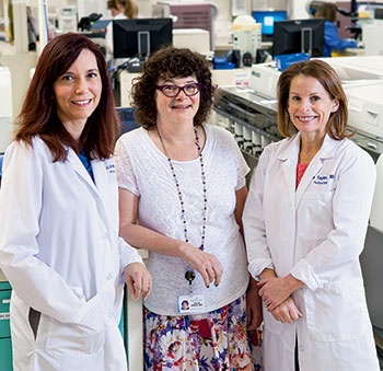
and oncologist Julie Asch, MD, associate director of the Intermountain Blood
and Marrow Transplant/Acute Leukemia Program. “Acute leukemias
are so diverse,” Dr. Cessna says, “and the testing seems to be exploding.”
“It’s hard for pathologists if every oncologist—and we have 30 or 40 here—wants something different,” says Rita Braziel, MD, professor, Department of Pathology, and director of the Hematopathology Division, Oregon Health & Science University, Portland.
Little wonder, then, that demand arose for a more formal guideline, addressing all acute leukemias; one is now being put together by an expert panel of CAP and American Society of Hematology members. The evidence-based guideline could be ready by early or mid-2015, says committee co-chair Daniel Arber, MD, the Ronald F. Dorfman, MBBch, FRCPath, professor of hematopathology, and vice chair of pathology for clinical services, Stanford University Medical Center.
But until then (and, frankly, even after), laboratories will have to continue their search for the best testing strategy. As the guideline committee and other pathologists know from experience, AML gives a decent imitation of perpetual motion. “It’s impossible to stop,” says Dr. Braziel. “Even if you take a standard subtype of AML, like acute promyelocytic leukemia [APL], which you think you know everything about—well, you don’t.”
During its literature search, the ASH/CAP guideline committee has discovered firsthand this unceasing aspect of AML, says Dr. Arber, who is also the medical director, anatomic pathology and clinical laboratory services, and co-associate director, molecular pathology laboratory. Recently Dr. Arber and his fellow co-chair, James Vardiman, MD, embarked on another round of data extraction from the articles (some 1,200 and counting). “That took quite awhile,” says Dr. Arber. “We didn’t have enough detail after the first round.” Another data refresh is likely at the end, he adds, acknowledging that newer data could likely support even stronger recommendations.
The initial push for the guideline came from Melissa Cessna, MD, a hematopathologist with Utah Pathology Services Inc. and Intermountain Healthcare, a community-based system. As Dr. Cessna observes, “Acute leukemias are so diverse, and the testing seems to be exploding. For community pathologists—and actually pathologists in any setting—understanding which tests should be done and how they’re used is important. But it’s hard to tease out.”
Dr. Cessna, who is on the committee, got her first taste of developing an AML testing algorithm when Intermountain expanded its bone marrow transplant service in 2006. The system now has a robust service for acute leukemia, she reports. Figuring out the right tests required working closely with the clinicians and balancing two competing but equally compelling demands: cost and choice. “I remember talking to one doctor who said, ‘I always like to have this marker,’” Dr. Cessna recalls. “She always had a ‘Did ya get?’ list—did you get this, did you get that?” Her reasons were sound. “You didn’t want to get stuck later not having a test that may have been relevant. But you were always guessing what might be relevant, and in that setting you would be shotgun ordering every test you might need.”
Intermountain’s strategy mimics air traffic by putting some tests in a holding pattern. Rather than ordering every FISH probe up front, Dr. Cessna and colleagues triage samples based on morphology and flow cytometry results. If they suspect APL AML, they then order stat FISH for t(15;17). Chromosome testing alone, she says, often provides much useful information. If the karyotype is complex, then the patient is known to have a poor prognosis, and additional FISH or molecular markers are unlikely to be relevant.

Molecular markers are “best proven only in certain situations,” Dr. Cessna continues. In patients with normal karyotype AML, FLT3, NPM1, and CEBPA will be prognostic. If FLT3 is positive, the prognosis will be poor. Mutated NPM1 indicates a good prognosis if there is no FLT3 mutation. Double-mutated CEBPA is associated with a good prognosis when FLT3 and NPM1 are negative.
Gone is the Old Country Buffet style of molecular testing in AML. Instead of laying out a spread, Intermountain now offers a series of tests, with each one requiring a clinical reason to move to the next “course.” “So for every new leukemia we just extract DNA and RNA, and there’s never a question of, ‘Did ya get this?’ Because we know we can add it on later,” says Dr. Cessna.
Doing so requires Dr. Cessna and colleagues to be in close contact with their reference laboratory as well as clinicians, however, and Dr. Cessna readily concedes Intermountain’s approach won’t work for everyone. Each lab, like Tolstoy’s unhappy families, is different.
Even in her own setting, Dr. Cessna sees the limitations. The hospital where she’s based serves a reference center, and it regularly sees patients who were previously diagnosed and perhaps treated with first-line induction therapy at another community hospital. Not all these patients have been tested for, say, FLT3, and they may not have had a DNA extraction. “We don’t have the capability to go back and add that, so you’re missing a piece of information that’s very important,” she says.
When Dr. Cessna and her colleagues created their algorithm, they soon identified that much of the acute leukemia testing was low volume. Even as a large community hospital, then, it would make sense to continue sending most of the molecular testing to an outside reference laboratory.
“We had a lot of discussion about what was practical,” Dr. Cessna recalls. “What was practical, and what we chose to employ, was to have our reference laboratory do DNA and RNA extractions.” In fact, they use two labs; one will hold material indefinitely, and the other for three weeks. “But we had to tell them what we needed, and they were willing to accommodate us.”
With AML specifically, she says, “We asked our reference laboratory, ‘What are the things that can be cryptic? What can be missed with a karyotype?’” In the resulting strategy, Dr. Cessna’s lab performs chromosome analysis, labeling it “high priority” so it can be triaged ahead of other cases. If the results are normal, it’s reflexed to three or four FISH probes known to alter prognosis, including inv(16), MLL gene rearrangement, and t(8;21). These results, in turn, will drive the molecular testing.
CEBPA is the most prognostic in the setting of a normal karyotype, negative FLT3, and negative NPM1 AML. “So if we have all those things, only then do we order the CEBPA,” Dr. Cessna says. KIT mutation testing is done only in cases with inv(16) or t(16;16) or t(8;21).
Having the lab hold material for possible later testing can slow turnaround times. Early on, Intermountain waited to order FLT3 until chromosome test results were available, but it now orders FLT3 at diagnosis, at the request of clinicians. Again, working closely with the reference laboratory paid off.
As for pushback from clinicians: “There was very little,” Dr. Cessna says. When the lab started looking into a new approach, it quickly became clear that many tests were being ordered unnecessarily, simply because oncologists were fearful of missing something. “But testing is expensive, and whether it’s relevant is dependent on what you see under the microscope,” Dr. Cessna says.
Intermountain offers so-called pathologist discretion for each type of test category. For flow cytometry, this is a “hold flow.” “We tell the lab whether or not to run it after we’ve reviewed the slides,” says Dr. Cessna. For cytogenetic studies, which include chromosome analysis and FISH, this will result in a “grow and hold,” where the laboratory will begin the cultures but not analyze them unless full analysis is requested. FISH can be added to a grow and hold or to a full chromosome analysis ordered up front within 90 days of collection. Grow and hold is a fraction the cost of the full study, she says.
“Adding FISH after you have the confirmatory diagnosis is also key,” Dr. Cessna says. “If you order based on what the diagnosis might be rather than what it is, there is a chance you’ll perform unnecessary or incorrect testing.” Take, for example, a patient who has pancytopenia, with AML in the differential diagnosis. If the AML panel is ordered up front, before the morphology has been reviewed, but the patient is later found to have metastatic breast cancer, then neither chromosome analysis nor FISH is needed. Ditto, she says, for flow cytometry—it would not be needed in this type of case, and it’s a very expensive test. “But if you order grow and hold, we can either not perform testing or add it after we’ve reviewed the slides. For molecular studies, we do a DNA and RNA extraction—the laboratory extracts the nucleic acids and stores them for us at -80°C.” She calls this approach “extract and store.”
In summary, she says, pathologist discretion results in (by category):
- flow cytometry—hold in flow cytometry laboratory (triaged next day or same day if Friday).
- cytogenetic studies (chromosome analysis, FISH)—grow and hold.
- molecular studies—DNA and RNA extraction. DNA-based tests include FLT3, NPM1, CEBPA, and mutations. RNA-based tests include RT-PCR for PML-RARA, etc.
Clinicians can order pathologist discretion for any or all of these, can order the testing up front to be run, or not order a test type (i.e. molecular).
“Most of the time, they let us triage these cases, especially if the diagnosis is unknown, except when they know they need chromosome analysis—that is, pre-BMT,” says Dr. Cessna. “What is nice about the grow and hold and DNA and RNA extraction is that the decision doesn’t have to be made right away because the material is held for so long.” That allows them to use their algorithm to drive which testing they perform, without compromising the specimens.
“Our clinicians like it. They like letting us decide what testing is indicated.”
“Personally, I like our strategy, too,” says Dr. Cessna, “because I know that material is there, and that I don’t have to go chasing a reserve sample from another test. And we’ve found that canceling testing already in progress is much more difficult than adding testing.”
“But other people like to perform it up front.”
Dr. Braziel, at OHSU, might be considered one of those “other people.”
“Our approach is to do complete testing up front for all new AML patients, so we know exactly what we’re dealing with,” she says. “And we find this information very helpful in follow-up.
“Our perspective is to invest in the complete characterization of a malignancy up front, then streamline follow-up testing and tailor it to the patient,” she continues. “That way, we don’t have any nasty surprises later in the course of disease in terms of additional poor prognosis mutations.” Even in AML cases with a good prognosis, additional genetic mutations can result in a bad outcome. “If you wait until a patient doesn’t do well, then your chance for definitive therapy may be compromised.”

Like Dr. Cessna, Dr. Braziel is familiar with the everyone-wants-something-different syndrome. Because OHSU is an academic institution, the “everyone” includes residents, fellows, nurse practitioners, junior faculty, and senior faculty, all of whom are involved in patient care. Until a couple of years ago, the lab was bombarded with calls from colleagues asking for this or that test. “It was so complicated, trying to train our hematopathology fellows and their hematology/oncology fellows, and figure out who wanted what to be done on whom,” Dr. Braziel recalls. That led to the hematopathology group meeting with the oncologists and, eventually, a standard institutional approach to working up patients with AML. The groups continue to meet intermittently and make changes as needed to adapt to new biologic discoveries. Their approach is a little more intensive than that of a community hospital, she says, because they’re involved in many clinical trials and testing new therapies.
“Now, [with] every new acute leukemia, the clinicians know certain tests will be done. They put in a standing order for acute leukemia testing, including flow cytometry, routine karyotyping, AML FISH, and molecular testing when the bone marrow is done,” says Dr. Braziel. “And since we often don’t know what kind of acute leukemia it is initially, they leave it up to us to order the appropriate FISH and the appropriate genetic testing based on what we see on flow and morphologic review.”
With AML patients at follow-up, she continues, “We also do a lot of flow cytometric sorting of relevant blast subpopulations.” She gives as an example a leukemic population with CD34 positivity and an inv(16) abnormality. If at follow-up there are only four percent CD34(+) blasts, which is within the normal marrow blast percentage, are they recovering normal blasts or residual leukemia? “Our clinicians want to know that,” she says. “They want to know the minimal residual disease burden.” In some cases, the standard FISH on the routine marrow aspirate is negative but the FISH on the sorted (>90 percent pure) CD34(+) myeloid blast population is positive. “We’re doing this type of testing all the time—paired flow cytometry to sort relevant subpopulations—and then doing FISH or molecular testing on that sorted population. For MRD and treatment planning, that’s very helpful.”
She works in an academic center with a particular interest in leukemia. “We do our own cytogenetics, FISH, and molecular testing,” she says. This includes a routine cytogenetics screen and a standard FISH panel for AML on every newly diagnosed acute leukemia case. They also perform next-generation sequencing on a panel of gene mutations—more than 40 at last count (see box), though the list fluctuates. “New AML patients come in at all stages of that workup.” Of seven cases that came in on a recent Friday, for example, three had already had blood and/or bone marrow work done before arriving at OHSU.
She and her colleagues at OHSU implemented the next-gen sequencing panel in response to the plethora of newly identified mutations of demonstrated or potential importance in AML. Standard, separate AML PCR analyses for the FLT3, NPM1, and CEBPA mutations, as well as KIT, were transferred to the NGS platform, which also includes other genes that are slowly entering the AML lexicon, such as JAK(1,2,3), IDH1, IDH2, DNMT3A, TET2, and RUNX1.
“Everybody has their favorite gene,” Dr. Braziel says, “and many new targeted therapies are being developed and tested relating to some of these genes.” The need to support individual investigators and clinical trials was a sticking point when the lab first broached with clinicians the topic of a more standardized approach to AML testing. Next-gen sequencing allows clinicians to include their favorite genes, without significant additional costs for testing. In another pleasing happenstance, reimbursement for the more standard mutations covers the costs of running the others. While it’s possible to run separate PCRs for FLT3, NPM1, and CEBPA, she says, “It ends up costing about as much as it does to do the entire NGS panel.”
Many acute myeloid leukemias can’t be adequately defined by those three mutations. “What if all three are negative?” Dr. Braziel asks. She says the NGS panel has been invaluable in this regard, as more than 90 percent of AML patients with a normal karyotype and FISH panel, as well as negativity for FLT3, NPM1, and CEBPA, have detectable mutations on NGS testing. About 50 percent of AML patients will have a normal karyotype, and many of those will have normal FISH results, too. That’s a substantial subgroup of patients who won’t initially have a detectable genetic abnormality. “NGS testing is a nimble platform for multiplex testing of multiple genes, including FLT3, NPM1, and CEBPA, in a single sample,” she says. It can be adapted quickly to delete genes later proved to be of little clinical relevance and to add new genes with potential predictive and therapeutic value.
Next-gen sequencing is also carving out a role for itself in myelodysplastic patients. Over the years Dr. Braziel and her colleagues have seen patients with cytopenias and multiple bone marrow evaluations in which cytogenetic and FISH tests have all been normal. “Often, there’s no firm diagnosis in these patients,” she says. In 50 to 60 percent of these patients, however, characteristic genetic mutations are present. “Our clinicians really appreciate having the molecular information on this subset of patients, who often go on to transplant.”
Two groups, two very different approaches, and two success stories. But some rough edges remain.
AML testing is complex and can fan out like Army Rangers on patrol. Dr. Cessna has seen cases where molecular tests have been sent to up to four different laboratories. The problem is not unusual when patients are diagnosed in one setting and receive treatment in another. But even when a patient remains within one system, problems arise.
Indiana University School of Medicine, Indi-anapolis, is the state’s only medical school, and “We consider ourselves the only academic health center in the state,” says Gail H. Vance, MD, professor of medical and molecular genetics and the Sutphin professor of cancer genetics. Certainly she and her colleagues have the resources to do AML testing onsite. “Although we’re all on the same campus, we test in different areas, which is not uncommon,” she says. “The hospital lab does the bone marrow aspirate; the pathology lab does the clot; I do the cytogenetics and FISH. And in a different lab, which I also direct, we do the CEBPA and the NPM1.” FLT3 is sent out—also a not uncommon practice, given some sticky patent issues with this mutation.
How do they coordinate all that information? “We make a lot of phone calls,” says Dr. Vance.
Stanford’s Dr. Arber says linking test results remains a big challenge at his institution. Final cytogenetic or molecular results often have to be added manually to reports containing results of bone marrow testing. It is, he says with understatement, “labor intensive, and it’s not very efficient.” Most laboratory computer systems aren’t yet up to the task, and in the interim, “A lot of people don’t want to integrate all the results into the report—they all come out at different times.”
“It’s cumbersome,” agrees Dr. Braziel, who says she and her OHSU colleagues also struggle to herd test results into one useful report.
These are the sorts of issues the ASH/CAP guideline committee hopes to address. Dr. Arber suspects there will likely be an expert recommendation to assemble the key information on a single report for the treating physician, though he concedes there are little data on the best way to do this.
And the matter is tied to a more complex question: Where should testing be performed? That, in fact, is the fifth of six questions the committee has set out to answer. It may also be the trickiest one, says Dr. Arber. “Should it be done in a community hospital?” he asks. “Or should they wait until the patient’s transferred to where they’re going to be treated [before] performing the testing?
“I don’t know that there’s a right answer,” continues Dr. Arber. “We haven’t found any literature yet that supports any answer,” which, given his experiences and those of Drs. Braziel, Cessna, and Vance, makes perfect sense. In clinical practice, the answers have been just as hard to find.
“Most people who are diagnosing acute leukemias don’t have immediate access to all of these molecular tests,” Dr. Arber notes. “So they’re going to have to send it somewhere.”
That launched passionate discussions on the committee. Some people were opposed to using a commercial laboratory for testing, arguing that the likelihood of errors increases when a test result is isolated from the rest of the case. (Dr. Braziel, who is not part of the committee, makes the same argument. “One of my concerns with national labs is specimen mixups,” she says. “We can pick that out when we get a result that’s anomalous and just doesn’t fit the rest of the case, and then we can do repeat testing. When you’re doing a test in isolation—you’re not seeing the blood, you’re not seeing the marrow—you’re not going to know if your result doesn’t fit.” When she and her colleagues have reviewed parallel results from OHSU and outside labs, she says, “We’ve had times where we’ve had some very clinically significant discrepancies.”)
Others argued that given the limitations of some community settings, it made sense to have all the testing done at the treating center. “But that’s also complicated,” Dr. Arber points out. “Because you don’t know that at the time you’re diagnosed,” and early on in the testing process, it may not even be clear that the patient has acute leukemia.
“And there’s a little bit of arrogance that only academic centers can do these things,” he says.
Given all this, Dr. Arber says he fully expects this section of the guideline, in its current draft form, to receive a rocky reception during the public comment period. The only bit sure to survive unscathed will be the portion recommending the testing be done in a CLIA-certified lab. “Which is the no-brainer part of it.”
Keeping materials at a single institution doesn’t guarantee ease, as Dr. Vance has noted. It’s also impossible to do if an institution sends out its FLT3 testing. This in turn creates more difficulties—physicians can be stymied when one mutation analysis arrives separately from the rest. For now, understanding the significance of an NPM1 result is impossible without knowing FLT3 status, but it’s quite likely other mutations will exhibit similar dependencies as well.
Again, these knotty clinical problems present a challenge for the guideline committee. In trying to answer the third question on its list—At the time of diagnosis, what tests are required for all patients for the initial evaluation of an acute leukemia?—they’ll need to take into account that the key physicians might well be people not yet involved in the case. So it’s not just a matter of what clinicians need—it’s what clinicians might need down the road, possibly at another center. “Know your referral pattern,” Dr. Cessna says. “Recognize that these centers may require testing that you didn’t perform.”
For the record, here are the other questions the committee will address:
What clinical and laboratory information should be available during the initial diagnostic evaluation of a patient with acute leukemia?
- What specimens and sample types should be evaluated during the initial workup of a patient with acute leukemia?
- Which tests should be performed only on a subset of patients, including in response to results of initial tests and morphology?
- How should test results and the diagnosis be correlated and reported?
Not on the list, but worth asking, is, How will the guideline fit into current practices?
Evidence-based answers will be welcome, observers say, since these are the crucial questions many labs are struggling with. Even those with algorithms could benefit from added insight.
It’s likely, says Dr. Braziel, that most academic institutions that regularly see acute leukemias have their own algorithms already, although she adds, “Frankly, it kind of depends on the personalities and whether people can work together and agree on a standard approach. At some places that doesn’t work, because everybody has their own little fiefdom.”
Even smaller institutions are likely to have a somewhat standardized approach, suggests Dr. Vance, given the growth in cancer consortiums. She sees many of the community hospitals in her area organizing—in Indianapolis alone, several local hospitals are affiliating with MD Anderson, she says, with which they share expertise and whose protocol they likely follow. “You can’t just look at it as an individual center anymore, or an individual pathologist,” she says. “You can look at individual academic health centers, but [they] also work in concert with one another, too.”
But unless the guideline is nimble, says Dr. Braziel, its usefulness will be limited. Dr. Arber shares that concern. “It’s a challenge to keep up with the literature. We don’t want this to be out of date when it appears.”
And for an evidence-based guideline, the evidence has, at times, been somewhat thin. Somewhat to Dr. Arber’s surprise, there was also very little evidence to firmly support additional molecular markers beyond the four most regularly used. “The data are quite variable,” he says. “I think we all tend to jump to conclusions—there will be one really good paper, and everyone will jump onboard, without looking at the other data. So this process has forced us to look at all the data.”
For now, the markers with the strongest basis for recommendation are CEBPA, FLT3, NPM1, and KIT. No surprise there. Others might be recommended, including IDH1 and IDH2, TET2, RUNX1, WT1, and DNMT3A. “But we’re still putting together data tables to support that,” Dr. Arber says.
At his own institution, he says, FLT3, CEBPA, and NPM1 testing is routinely done for all AMLs; KIT is added in cases with a core binding factor translocation. The lab is also getting ready to go live with an extended myeloid mutation panel.
Intermountain hasn’t taken the next-gen leap yet, but it will likely do so in the next year, Dr. Cessna says. She worries that more information, if not used wisely, could simply be more information. She’s not alone in her concern. Before Intermountain established its current algorithm, she says, the laboratory used to do considerably more follow-up testing than it does now. The lab eventually cut back when clinicians said they didn’t want additional testing—they didn’t know what to do with the results, and the tests only made the patients nervous, says Dr. Cessna. “More information doesn’t always make things clearer.”
The guideline won’t explicitly address next-generation sequencing, but it’s possible next-generation sequencing will eventually take on the guideline. “We’ve been working through all this literature, trying to come up with these algorithms, and now next-generation is on the scene—and it could change everything,” says Dr. Cessna.
[hr]
Karen Titus is CAP TODAY contributing editor and co-managing editor. Dr. Vance suggests two resources for pathologists. One is an Archives of Pathology & Laboratory Medicine article, “Acute myeloid leukemia diagnosis in the 21st century” (Betz BL, et al. 2010;134:1427–1433). The other is the 2012 CAP webinar on AML diagnosis, presented by David Czuchlewski, MD, and Mohammad Vasef, MD (http://j.mp/amldx—CAP user ID and password needed for access). Dr. Arber will present a session with Dr. Vardiman at CAP ’14 on Sept. 10 on the CAP/ASH Algorithm for Initial Workup for Acute Leukemia.