CAP TODAY and the Association for Molecular Pathology have teamed up to bring molecular case reports to CAP TODAY readers. AMP members write the reports using clinical cases from their own practices that show molecular testing’s important role in diagnosis, prognosis, and treatment. The following report comes from the University of Minnesota. If you would like to submit a case report, please send an email to the AMP at amp@amp.org. For more information about the AMP and all previously published case reports, visit www.amp.org.
Guang Yang, MD, PhD; Michelle Dolan, MD
Haixia Qin, MD, PhD; Manish R. Patel, DO
Sophia Yohe, MD; Andrew C. Nelson, MD, PhD
July 2021—The advent of genomically targeted therapy and immunotherapy has greatly altered the clinical management of advanced non-small cell lung cancer (NSCLC).1 Molecular testing is recommended for sensitizing EGFR mutations, ALK fusions, ROS1 fusions, BRAF V600E, NTRK fusions, RET fusions, and MET exon 14 skipping alterations.2 Anaplastic lymphoma kinase (ALK) gene rearrangements are identified in about three to seven percent of NSCLC cases, with the echinoderm microtubule-associated protein-like 4 (EML4) gene being the most common fusion partner.3
Multiple small molecule inhibitors targeting ALK fusion proteins have been developed, and the use of these new therapies has significantly improved the overall survival of patients with ALK-rearranged NSCLC.4 However, as with many targeted therapies, eventual resistance to ALK inhibitors and disease progression are often observed in NSCLC patients.5,6 Primary resistance refers to a lack of tumor response from the initiation of the targeted therapy, whereas secondary resistance is defined as disease progression after an initial partial or complete response to treatment.7 Although most studies focus on various secondary-resistance mechanisms along with therapeutic strategies, the molecular mechanisms of primary resistance to ALK inhibitors in ALK-rearranged NSCLC are not well documented.8
We report a case with primary resistance to ALK inhibitors in EML4-ALK-positive lung adenocarcinoma with concomitant MYC amplification. We also review the related literature and discuss possible therapeutic strategies to overcome this primary resistance to ALK inhibitors.
Case presentation. A 68-year-old Caucasian female nonsmoker presented with a two-month history of dry cough, shortness of breath, worsening mid-thoracic back pain, and new onset mid-sternal chest pain. Chest computed tomography pulmonary angiogram with contrast showed two 10–12-mm solid pulmonary nodules in the right upper lobe. A positron emission tomography scan of the neck, chest, abdomen, and pelvis demonstrated widespread metastases in bones, liver, adrenal, and retroperitoneal lymph nodes. Magnetic resonance imaging of the brain detected numerous punctate brain metastases.

Fig. 1. Endobronchial ultrasound-guided fine-needle aspiration of station 10L lymph node. H&E 20 ×: variable-sized fragments of tumor cells (upper image). TTF1 20 ×: tumor cells show weak, patchy nuclear positive for TTF-1 (lower image).
Endobronchial ultrasound-guided fine-needle aspiration of station 10L lymph node showed non-small cell carcinoma. Tumor cells demonstrated weak, patchy nuclear positivity for TTF-1 and were negative for p40. Based on these findings, a diagnosis of adenocarcinoma of pulmonary origin was made (Fig. 1). PD-L1 immunohistochemistry (Ventana clone SP263) showed low expression (tumor proportion score between one and two percent).
DNA-based next-generation sequencing was performed using a custom-designed targeted 61-gene hotspot panel, which covers single nucleotide variants (SNV) and small insertions/deletions (indel). Library preparation was carried out by tagmentation following Nextera protocols (Nextera XT library preparation, Illumina). The enriched libraries were sequenced on an Illumina MiSeq instrument to a target of 1.5 million reads per sample. No actionable SNV or small indel were detected in the genes sequenced (ALK, BRAF, EGFR, ERBB2, IDH1, IDH2, KRAS, MET, NRAS, PIK3CA, RET, and TP53).
A parallel RNA-based targeted NGS assay was performed for the simultaneous assessment of fusions, exon skipping, and other expression targets frequently observed in NSCLC. Targeted RNA-Seq libraries were prepared using the Quantidex NGS RNA Lung Cancer Kit (Asuragen) according to the manufacturer’s instructions. EML4-ALK gene rearrangement (EML4 exon 20 fused to ALK exon 20) was detected. Based on this finding, the patient was treated with alectinib, a highly potent second-generation ALK-specific kinase inhibitor, 600 mg twice daily. No conventional chemotherapy was used.
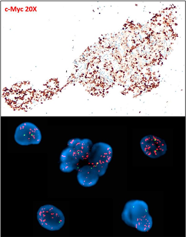
The patient’s clinical symptoms were initially stable, but within one month she developed headaches and had evidence of progression by restaging MRI, which showed many new enhancing intracranial lesions. Guardant360 74-gene cell-free DNA (cfDNA) NGS assay (Guardant Health) with a clinical sensitivity of 85 percent9 was performed on peripheral blood specimen and identified EML4-ALK fusion (11.2 percent cfDNA). This assay also showed the unexpected finding of high-level MYC amplification with an estimated plasma copy number of 33.9 (normal copy number=2).10 No ALK mutations associated with resistance to inhibitors, or bypass mutations in other pathway genes, were identified. Immunohistochemical analysis of c-Myc (clone Y69 from Abcam) performed on the lymph node fine-needle aspiration specimen obtained at diagnosis (i.e. before therapy) demonstrated strong diffuse nuclear MYC positivity in most of the neoplastic cells (Fig. 2, upper image). Fluorescence in situ hybridization on the same diagnostic material using a breakapart probe to the MYC locus (Abbott Laboratories) showed amplification of the 5’ (centromeric) portion of the probe (Fig. 2, lower image).
Because of the symptomatic progression of the patient’s multiple brain metastases, the patient was treated with steroids and later with palliative whole brain radiotherapy. Several days later she was admitted with sepsis due to suspected aspiration pneumonitis, postobstructive pneumonia, and right-sided pleural effusion. After admission the patient was started on antibiotics, but her overall condition rapidly deteriorated and she died of acute respiratory failure three months after starting targeted therapy.
Discussion. Over half of newly diagnosed patients with lung cancer have metastatic disease at the time of diagnosis, making surgical intervention ineffective.11 Therapy targeted at the underlying molecular abnormality has significantly improved the clinical outcomes of subgroups of NSCLC patients with actionable mutations in EGFR, ALK, ROS1, RET, BRAF V600E, MET exon 14, and NTRK.12 Multiple ALK tyrosine kinase inhibitors, including crizotinib, ceritinib, alectinib, brigatinib, and lorlatinib, have been approved by the Food and Drug Administration for the treatment of patients with advanced ALK-positive NSCLC.13
Among these TKIs, alectinib is currently considered the preferred first-line treatment because of its clinical activity and favorable toxicity profile.2 According to a recent study, alectinib is more effective than standard chemotherapy in patients with advanced or metastatic ALK-positive NSCLC, with a median progression-free survival significantly longer with alectinib than with chemotherapy (9.6 months versus 1.4 months).14 Studies have also demonstrated that in patients with advanced ALK-rearranged NSCLC, alectinib has a higher brain penetration15 and overall central nervous system response rate15 than crizotinib (85.7 percent versus 71.4 percent, respectively).
Although the use of ALK TKIs has led to marked improvements in response and survival, patients with ALK-rearranged NSCLC inevitably develop secondary resistance.16 Current research on resistance to ALK inhibitors in NSCLC largely focuses on identifying and characterizing the molecular mechanisms causing secondary resistance, such as mutations or amplification of ALK and “bypass track” activation via mutations in EGFR and PIK3CA, amplification of MET and KIT, and IGF1R activation.7 Secondary mutations in the ALK kinase domain were observed in about 50 percent of the ALK-rearranged NSCLC cases with resistance to second-generation ALK TKIs.17 In a study of 51 ALK-positive NSCLC patients who had progressive disease upon treatment with crizotinib, the most common ALK resistance mutations were L1196M and G1269A, detected in seven percent and four percent of these patients, respectively. Other mutations identified were C1156Y (two percent), G1202R (two percent), I1171T (two percent), S1206Y (two percent), and E1210K (two percent).18 However, mechanisms of primary resistance to ALK inhibitors are less well characterized.
Our patient was treated with a second-generation ALK inhibitor, alectinib, shortly after her diagnosis of metastatic EML4-ALK-positive lung adenocarcinoma. Her tumor showed no objective response to this targeted therapy, with radiologic evidence of progression within one month of therapy initiation. Because the median overall survival for stage III/IV ALK-positive NSCLC patients treated with alectinib is 48.2 months,19 this patient’s rapid deterioration and death after ALK targeted therapy was unexpected. In light of the rapid disease progression while on alectinib, we hypothesized that this patient’s aggressive clinical course was likely due to a primary resistance mechanism not characterized in the guideline-focused genomic assays performed at diagnosis.

Fig. 2. Endobronchial ultrasound-guided fine-needle aspiration of station 10L lymph node. c-Myc 20 ×: tumor cells show strong nuclear positive for c-Myc (clone Y69 from Abcam) (upper image). FISH using a dual-color breakapart probe to the MYC locus (Abbott Laboratories) shows amplification of the 5′ (orange, centromeric) portion of the probe; two signals for the green (3′ telomeric) probe (lower image).
The cfDNA assay performed at the time of progression identified high-level MYC amplification, which was retrospectively confirmed to be present at diagnosis by IHC and FISH studies performed on pretreatment specimens.
 CAP TODAY Pathology/Laboratory Medicine/Laboratory Management
