
Collection and handling takeaways for cytopathology practice
Sinchita Roy-Chowdhuri, MD, PhD
August 2020—Cytopathologists are keenly aware of the need to collect adequate cytologic tissue not only to arrive at a diagnosis but also to provide sufficient material for predictive and prognostic markers. This is especially true in the realm of non-small cell lung cancer, where biomarker testing is routinely used for the clinical management of patients with advanced-stage disease. The list of clinically relevant biomarkers in NSCLC is expanding.
The most recent version of the National Comprehensive Cancer Network Clinical Practice Guidelines in Oncology includes MET exon 14 skipping mutations and RET as therapeutic targets for advanced NSCLC, in addition to the well-established EGFR and BRAF mutations, ALK and ROS1 rearrangements, and PD-L1 expression.1 Testing modalities for these seven “must test” biomarkers is extensive, ranging from PCR-based methods for mutational analysis to fluorescence in situ hybridization assays to identify gene rearrangements, to immunohistochemistry for quantifying protein expression in tumor cells. Not surprisingly, small specimens collected by minimally invasive techniques, such as fine-needle aspiration and core needle biopsy, often fall short in meeting adequacy requirements for the myriad testing modalities for a growing list of biomarkers.2,3
The CAP has been at the forefront in leading laboratory practice by developing evidence-based practice guidelines that help direct clinicians and laboratories. To that end, the CAP recently published the guideline on collecting and handling thoracic small biopsy and cytology specimens for ancillary studies, which is aimed at providing direction to proceduralists and pathology laboratory personnel for the most optimal collection and handling of such specimens.4 The guideline was developed in collaboration with stakeholders from eight other professional medical societies: American College of Chest Physicians, American Society of Cytopathology, American Thoracic Society, Association for Molecular Pathology, Papanicolaou Society of Cytopathology, Pulmonary Pathology Society, Society of Interventional Radiology, and Society of Thoracic Radiology. A multidisciplinary expert panel, composed of cytopathologists, a cytotechnologist, molecular pathologists, pulmonary pathologists, interventional pulmonologists, interventional radiologists, and a research methodologist, with input from a separate advisory panel, worked through 4,256 peer-reviewed published studies that were identified in the systematic review to develop 16 guideline statements on the best practices for acquiring and handling thoracic small specimens for ancillary studies.
The guideline covers six main aspects of thoracic small specimen acquisition: endobronchial ultrasound-guided transbronchial procedures, transthoracic procedures, bronchoscopic procedures, pleural effusions: considerations for malignancy, considerations for ancillary studies during malignant investigations, and considerations for ancillary studies during nonmalignant investigations. It includes a range of ancillary studies, from those used for patients with lung cancer to those used in the diagnosis of infectious processes such as pulmonary and extra-pulmonary tuberculosis. While a detailed description of the entire guideline is beyond the scope of this article and the reader is directed to the published guideline in the Archives of Pathology & Laboratory Medicine, some of the guideline statements are of particular importance to the cytopathology community, and a brief overview is provided (Table 1).
Keeping in mind that there are existing recommendations from other professional medical societies pertaining to procedural aspects,5,6 the aim of the new guideline is to provide direction for collecting a thoracic small specimen that is adequate for downstream ancillary testing. With respect to cytology, specific guidelines are provided to the proceduralist pertaining to the choice of needle gauge (statement Nos. 2 and 6), number of passes (statement Nos. 4 and 7), utility of rapid on-site evaluation (ROSE) (statement Nos. 3, 4, 5, and 10), and optimal volume and triage of pleural effusion specimens (statement No. 11).
The multiple guideline statements pertaining to the use of ROSE, when available and clinically feasible, highlight the role of the cytologist during minimally invasive procedures, a role that can ensure adequate sampling of the targeted lesion as well as appropriate specimen triage for necessary ancillary studies. While overall the use of ROSE is recommended whenever possible, the expert panel acknowledges the potential complexity of different practice settings and clinical scenarios in which ROSE may not be needed or even practical, leaving room for clinical judgment to be used in determining the need for ROSE in all procedures.
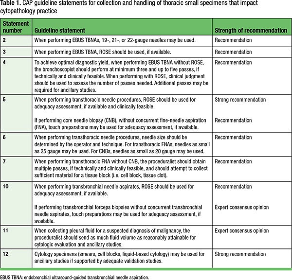
Similarly, the expert panel suggests sending as much fluid volume as can be reasonably attained to the cytopathology laboratory for pleural effusions that are collected with a clinical suspicion of malignancy. This guideline statement comes as an expert consensus opinion because there was insufficient evidence in the systematic literature review to recommend a specific effusion volume that would be considered adequate for ancillary studies. The general assumption is the adequacy of a pleural effusion depends on the cellularity of the fluid, and therefore the larger the volume submitted to the laboratory, the greater the chances the specimen would be adequate for ancillary studies.
An additional guideline statement (No. 12) of particular importance to the cytopathology community is the strong recommendation for the use of all cytology specimens, i.e. smears and liquid-based cytology in addition to cell blocks for ancillary studies, when supported by validation studies. This is of special relevance because historically most ancillary studies in cytology, including molecular/PCR-based testing, FISH, and IHC, have primarily been limited to formalin-fixed, paraffin-embedded cell block preparations due to their similarity to histologic tissue blocks. The use of non-FFPE cytologic substrates requires additional validation studies that pose a major limitation to their widespread use, especially in reference laboratory and commercial settings.7-10 However, with the growing number of ancillary biomarker studies required for NSCLC, it is important for the cytology and molecular community to recognize the potential advantage of using the various cytologic preparations judiciously to be able to provide adequate testing needed for patient care. This has been highlighted in the most recent iteration of the CAP/ International Association for the Study of Lung Cancer/ Association for Molecular Pathology molecular testing guideline for lung cancer, where the use of all cytologic preparations was recommended in contrast to the prior preference for cell blocks.11
In conclusion, this new guideline provides direction to pathologists, laboratory personnel, and our clinical colleagues for better collection and handling of thoracic small specimens that are adequate for ancillary studies to help guide therapeutic decisions. However, one of the major limitations highlighted in the guideline is the paucity of high-quality, well-designed published studies in cytology that address some of the preanalytic variables the expert panel had hoped to address in this guideline. For instance, there was inadequate data in the systematic literature review to warrant a recommendation for the choice of a collection medium, fixative, or stain in cytology specimens for ancillary studies and insufficient data to provide guidance for recommended cold ischemic time or duration of fixation for optimal ancillary testing success. This underscores a need for high-quality published studies in cytology that provide comparisons of preanalytic variables between specimen preparations, testing methods, and practice settings to provide better granularity and guide future guideline efforts. This is important for the cytology community to convey broadly, as we continue to move toward using minimally invasive sampling techniques for a growing number of ancillary studies, thus putting these small specimens at the forefront of precision medicine. The CAP and the eight collaborating medical societies that approved the guideline are encouraging all of their members to adopt the guideline recommendations and coordinate efforts to determine how best to implement these recommendations in their clinical practice.
1. National Comprehensive Cancer Network. NCCN Clinical Practice Guidelines in Oncology: Non-Small Cell Lung Cancer. Version 5.2020. Accessed June 1, 2020. www.nccn.org/professionals/physician_gls/pdf/nscl.pdf.
2. Arcila ME, Oxnard GR, Nafa K, et al. Rebiopsy of lung cancer patients with acquired resistance to EGFR inhibitors and enhanced detection of the T790M mutation using a locked nucleic acid-based assay. Clin Cancer Res. 2011;17(5):1169–1180.
3. Sabir SH, Krishnamurthy S, Gupta S, et al. Characteristics of percutaneous core biopsies adequate for next generation genomic sequencing. PLoS One. 2017;12(12):e0189651.
4. Roy-Chowdhuri S, Dacic S, Ghofrani M, et al. Collection and handling of thoracic small biopsy and cytology specimens for ancillary studies: guideline from the College of American Pathologists in collaboration with the American College of Chest Physicians, Association for Molecular Pathology, American Society of Cytopathology, American Thoracic Society, Pulmonary Pathology Society, Papanicolaou Society of Cytopathology, Society of Interventional Radiology, and Society of Thoracic Radiology. Arch Pathol Lab Med. Epub ahead of print May 13, 2020. doi:10.5858/arpa.2020-0119-CP.
5. Sokolowski JW Jr, Burgher LW, Jones FL Jr, Patterson JR, Selecky PA. Guidelines for percutaneous transthoracic needle biopsy. This position paper of the American Thoracic Society was adopted by the ATS Board of Directors, June 1988. Am Rev Respir Dis. 1989;140(1):255–256.
6. van der Heijden EHFM, Casal RF, Trisolini R, et al. Guideline for the acquisition and preparation of conventional and endobronchial ultrasound-guided transbronchial needle aspiration specimens for the diagnosis and molecular testing of patients with known or suspected lung cancer. Respiration. 2014;88(6):500–517.
7. Roy-Chowdhuri S, Chen H, Singh RR, et al. Concurrent fine needle aspirations and core needle biopsies: a comparative study of substrates for next-generation sequencing in solid organ malignancies. Mod Pathol. 2017;30(4):499–508.
8. Roh MH. The utilization of cytologic fine-needle aspirates of lung cancer for molecular diagnostic testing. J Pathol Transl Med. 2015;49(4):300–309.
9. da Cunha Santos G, Saieg MA. Preanalytic specimen triage: smears, cell blocks, cytospin preparations, transport media, and cytobanking. Cancer Cytopathol. 2017;125(S6):455–464.
10. Roy-Chowdhuri S, Aisner DL, Allen TC, et al. Biomarker testing in lung carcinoma cytology specimens: a perspective from members of the Pulmonary Pathology Society. Arch Pathol Lab Med. 2016;140(11):1267–1272.
11. Lindeman NI, Cagle PT, Aisner DL, et al. Updated molecular testing guideline for the selection of lung cancer patients for treatment with targeted tyrosine kinase inhibitors: guideline from the College of American Pathologists, the International Association for the Study of Lung Cancer, and the Association for Molecular Pathology. Arch Pathol Lab Med. 2018;142(3):321–346.
Dr. Roy-Chowdhuri is an associate professor and molecular diagnostic laboratory director (solid tumors) at the University of Texas MD Anderson Cancer Center, Houston. She practices cytopathology and molecular pathology. She is a member of the CAP Cytopathology Committee and was co-chair for the CAP Guideline for Collection and Handling of Thoracic Small Biopsy and Cytology Specimens for Ancillary Studies.