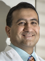
Dr. Compton
Dr. Compton notes that variations in the fixative, for example, can affect the downstream analyses of these tissues. “What we want is a standard fixative, 10 percent neutral phosphate-buffered formalin, that is carefully quality controlled, because it preserves both morphology and a whole range of biomolecules quite well. This ties to the digital piece because if you use alcohol fixative, for example, which fixes by dehydration and denaturation in contrast to formalin, which fixes by cross-linking of molecules, the results will be different. Alcohol causes subtle changes in the nuclear morphology because it causes DNA to collapse, as well as cytoplasmic changes because it produces protein coagulation and cell shrinkage. But if you have a rock solid standard against which your digital technology can do quantitative measurements, you’d be able to detect samples that would be inappropriate for analysis because of preanalytical factors.”
Total time in formalin must also be standardized. “No less than six hours and no more than 24 to 36 hours. Improper fixation time can result in either under-fixation with inadequate molecular preservation or over-fixation with molecular damage,” Dr. Compton says, “even artifactual mutations in the DNA. The fallout could be tremendous: the wrong answer on a molecular analysis.”
Dr. Compton believes digital pathology may hold the key to improving specimens. “Some of the preanalytical steps cause artifacts in tissue that may be difficult or impossible to detect by the human eye on a glass slide. But if digital pathology were to use its power to detect subtle changes in morphology or tintorial properties and compare those to a standard, it could then flag specimens that are unreliable for biomarker staining, detection, or analysis and alert us that they have been compromised in the preanalytical phase,” she says. “That would be a huge leap forward.”
Dr. Compton says pathology is just beginning to uncover the myriad benefits within digital technology. “I don’t know exactly where digital pathology will fit in,” she concedes, “but I do think it is a more precise tool to determine specimen acceptability than just doing it by the seat of our pants. We are still very early in the discovery mode with respect to digital pathology. Its potential is not yet fully unlocked.”
Others agree with that premise of expanding utility. Dr. Glassy says digital imaging brings into play additional quality control supports, ones that technologists are accustomed to in clinical labs. “Tools, graphs, and Westgard rules can now be implemented within histology. Whereas before histotechs did not have a good measurement of the quality of their stains, digital imaging allows that to happen.”
A laboratory can establish and install rules and algorithms within its technology to flag something out of range. “That kind of scientific rigor is being applied to what was before a purely manual analog process. It allows us to quantitate how things have changed. Are your sections too thick? Are your immunohistochemical stains still at the same intensity they were a month ago when you first opened the lot of reagents? These things are not easily determined by a histotechnologist or a pathologist who is looking at a slide. But a digital scanner can measure the intensity of the staining from day to day, and that can be plotted out on a graph. Some labs are doing this now. It’s easy to implement,” Dr. Glassy says.
Optimizing such capabilities will require training that emphasizes workflow adjustments and observance of quality control.
“Workers will need to understand required changes to the workflow; that is part of the frozen section story in diagnosis,” Dr. Laszik says. “You cut the section, you coverslip them, and in the old model you delivered that to the pathologists who were sitting nearby and looked at the slides. Now you need to scan them, and what if scanning goes wrong? What are the backup solutions? How many times do you need to rescan those slides? If you cannot get it in focus, do you need to cut new sections?” These are some of the issues that should be addressed from a workflow standpoint, he says.
While its early track record is impressive, digital pathology is not a perfect science, and laboratories must be prepared for the imperfections that may arise, albeit infrequently. Digital pathology was introduced in Dr. Laszik’s lab in early 2015 for frozen sections, and he and colleagues devised a backup solution in the event something were to go awry. “We have three hospitals and all three have ongoing operating rooms with frozen sections, scanners. We realized we could use one person instead of three to read frozen sections,” he says. “But we also had the luxury of an on-site pathologist who could cover in case of a problem with the scanner or a bad scan. Yet I can say that since we started to do these in 2015 there was not a single case out of hundreds of cases that required something that would be extraordinary. There were, however, a few cases that required an on-site look at the case. So you must be prepared for even one case.” If there is no pathologist on site, one alternative backup solution could be a live WebEx session, he suggests.
“The only way to make people aware of best practices for specimen and slide preparation and troubleshooting the unexpected that may arise is training, training, and more training,” Dr. Laszik emphasizes. “Train your people, monitor the quality all the time, and give them feedback. It is an old-fashioned approach, but it is even more significant in the digital age than it was before.”

Dr. Parwani
Dr. Parwani, too, sees greater QC potential in the technology that drives digital pathology. “Digital pathology is an enabling technology,” he says. “Until now there was no objectivity; pathology was an art rather than a science. But now we have measurable levels of standardization that we can take advantage of. We can run a computer program with a lab’s own rules for an acceptable quality threshold. We can add a number to it and give an image a quality score. Any image that does not meet a predetermined measure fails. Furthermore, we can review images that come out of the histology lab and provide instantaneous feedback to histotechnologists. This creates a dynamic feedback loop into the lab.” When slides are digitized, he says, true collaboration is possible. “This is the beauty of digital slides: We can manage workflow and make everything more streamlined in terms of image management, image sharing, and image analysis—three things we could not do with glass slides.”
Laboratory staff can be educated and processes improved at the same time, Dr. Parwani says. “That is very, very powerful.”
The associated utilities of digital pathology, bolstered by underlying, high-quality histotechnology, seem to have no observable limit on the horizon. “It’s like a pyramid,” Dr. Parwani says. “We are at the very bottom of the pyramid now where we are seeing more and more labs starting to digitize their slides. Once the base of the pyramid is filled with images, we can build on it. We can expand in the areas of research, companion diagnostics, quality assurance, education, consultations. The sky is the limit.”
[hr]For histotechnologists and pathologists, a Digital Pathology Certificate Program
A new Digital Pathology Certificate Program, a collaborative effort of the National Society for Histotechnology and the Digital Pathology Association, is expected to roll out this fall.
“NSH realizes that digital pathology is becoming more important and that the tasks of keeping scanners working optimally, maintaining proper validations, etc., generally falls to histotechs,” DPA president Eric Glassy, MD, says. “NSH hopes to recognize the extra interest and hard work their members are providing in support of digital pathology, and wants them to be recognized as imaging technologist specialists.”
The course is suitable for others who might want to achieve the same competence. “This is not just histotech focused. It is for pathologists as well, who may wish to be certified in proper use of whole slide imaging scanners,” Dr. Glassy says.
To earn the certificate, participants in the program will complete seven modules: introduction and history of digital pathology; basics of the technology—hardware/software; use cases; implementing and selecting a digital pathology solution; workflow considerations/best practices; image analysis; and regulatory/validation.
The course will be the topic of a panel discussion at the DPA’s Pathology Visions meeting in San Diego, Oct. 1–3.
Also noteworthy among training efforts, says Dr. Glassy, is a forthcoming module within the CAP/NSH HistoQIP program that will allow the grading of a digital scan. “The hope is to unveil it early next year,” he says. “Right now criteria for how scans should be evaluated and properly graded are being developed by two CAP committees. Eventually there will be two types of proficiency testing—one a virtual HistoQIP program and the other an evaluation of the scan.”
For histotechnologists, this will serve to validate they are performing to a high standard, Dr. Glassy says.
“And this will really expand the reach of the HistoQIP programs. This is a worldwide program; other countries may submit slides for evaluation.” The reach of digital pathology has already taken existing HistoQIP modules to countries that formerly could not participate—China, for one. “No human tissue may leave the country, so Chinese lab professionals can’t send out paraffin blocks or glass slides,” Dr. Glassy says. “But because DP scanning centers have been set up, HistoQIP can now move into countries that limit how slides can be sent for grading.” —Valerie Neff Newitt
[hr]Valerie Neff Newitt is a writer in Audubon, Pa.
 CAP TODAY Pathology/Laboratory Medicine/Laboratory Management
