Anne Paxton
October 2023—In living up to its promise as a new technology that will revolutionize clinical care through greater ease, speed, and accuracy of diagnosis, digital pathology has been sluggish. While many analysts, starting at least two decades ago, forecasted that digital pathology would elbow aside glass slides for good, that milestone is still far out of reach.
As health economist and chief executive officer of the New York City-based digital pathology company Paige, Andy Moye, PhD, puts it bluntly: “In probably 90 to 95 percent of the cases in the U.S., a pathologist still makes the diagnosis of cancer the way they did it back in 1910: by looking at a glass slide under a microscope.”
Mark Lloyd, PhD, vice president of pathology for Fujifilm, says he wouldn’t be surprised to hear that perhaps only five percent to 10 percent of hospitals have moved beyond using only glass slides to offer pathologists digital pathology capability. In fact, Dr. Lloyd thinks those percentages are overstated. What is the market share for the clinical use of digital pathology? “I think it would be generous to say it’s five percent. In the U.S. specifically, digital sign-out is extraordinarily low.”
However, he views Fujifilm’s purchase last January of the digital pathology business from Inspirata as an important signal, among others, that digital pathology’s star is rising. Fujifilm, a $26 billion company with 80,000 employees and already highly experienced in digital radiology, “believes the needle is moving. They’ve voted on digital pathology as being that next major digital transformation.” This indicates maturity of digital pathology in the clinical market, he says.
There are signs that, in line with Dr. Lloyd’s belief that the needle is moving, digital pathology is doing more than occupying a niche in the clinical diagnostics landscape:
- Two years ago, the FDA approved for marketing the first product to combine digital pathology with artificial intelligence, Paige’s Prostate Detect. In June and July of this year, Paige launched its suite of breast and colon cancer digital pathology products for which the company is seeking approval.
- Paige announced last month it would collaborate with Microsoft to build “the world’s largest image-based artificial intelligence models for digital pathology and oncology.” Configured with billions of parameters and incorporating up to 4 million digitized microscopy slides across multiple types of cancer, the new AI model is “orders of magnitude larger than any other image-based AI model existing today,” the company says.
- Fujifilm’s new Synapse Pathology PACS, a “vendor agnostic” software, delivers digital images for diagnosis 1.7 hours faster than glass slides, the company says, and is the only such software with “substantially equivalent” 510(k) FDA clearance with multiple scanners to market in the United States. Designed for large medical facilities across multiple locations and compatible with the products of multiple scanning vendors, Synapse Pathology became available in March this year.
- On the adoption front, Mayo Clinic is starting the third phase of a several-year digital pathology initiative, the aim of which is to replace glass slides for clinical diagnosis. “They’re setting up an entire platform, a kind of marketplace where they can collaborate and where all different kinds of digital data can be shared and maybe where you could create, for example, algorithms based on AI,” says Esther Abels, MSc, a precision medicine and biomedical regulatory health science expert and former president of the Digital Pathology Association.
Partnerships among digital pathology companies, facilitating streamlined digital workflow between scanners, image management software, and artificial intelligence tools, are proliferating, DeciBio Consulting says.
In its “Digital and Computational Pathology Market Report—First Edition: 2023–2028,” released this May, the company reports that about 40 digital pathology partnerships were announced in the first four months of this year.
DeciBio views partnerships as key to ensuring access to digital pathology capabilities in the clinical setting. Senior DeciBio product manager Katie Gillette cites the example of Fujifilm, which, with its acquisition of Inspirata, now has the largest number of partnerships in the digital pathology space as the market continues to evolve to better address customers’ needs. Other digital pathology companies, such as PathAI, Paige, Proscia, Mindpeak, Nucleai, Owkin, and Visiopharm, have also made partnerships for interoperability and research a priority, she adds.
DeciBio estimates the worldwide clinical digital pathology market in 2023 at about $270 million and growth of 15 percent per year, reaching $550 million in 2028. The U.S. is estimated to account for about 40 percent of that and to grow in line with the market.
In the report, projected growth in the U.S. market is broken out by customer segments. Within academic medical centers, where clinical use of digital pathology is most advanced, expected growth is close to 12 percent per year. In community hospitals, which are a much smaller segment of the market today, higher growth of 15 percent per year is projected, driven by initial adoption of scanners and image management systems in community hospital systems that don’t have the infrastructure now, Gillette says.
“Based on the feedback from the academic medical centers, AI tools for primary diagnosis and therapy selection are expected to be key growth drivers,” she says.

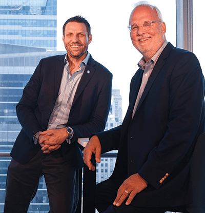
Andy Moye, PhD (left), chief executive officer of Paige, with David Klimstra, MD, Paige co-founder and chief medical officer. “At the end of the day, we believe this field is sort of the last frontier in digitization,” Dr. Moye says of pathology. [Photo by Jennifer Altman]
Measuring digital pathology’s adoption in U.S. hospitals is difficult, Gillette says, because there is no consensus on what “all digital” means. “Even in some of the institutions that are most bullish on digital pathology, there are still specific pathologists, labs, or subspecialties that do some or all of their cases manually.” They may say, for example, “‘Today, most of our sign-outs are digital’” or refer to efforts to establish a fully digital workflow without specifically indicating which parts of their workflows and which slides are digitized, she says.
Based on survey results, primary interviews, and secondary data, DeciBio estimates that about 10 percent of academic medical centers and less than one percent of community hospitals currently use digital pathology to support routine primary diagnosis.
A digital pathology workflow has multiple components, Gillette notes. “You need the slide scanner hardware, then you need the image management software to be able to view and annotate work and manage a patient’s case. You may use image analysis tools, and you may also need storage in the cloud.”
“There’s the whole digital pathology ecosystem that exists outside of that. And it takes a lot of change management and implementation to get digital pathology up and running.”
Many of the clinical lab respondents to DeciBio’s survey said they had scanners, for example. “But when you dive into it,” Gillette says, “they may have the scanner but are only scanning the most confusing cases, the cases that needed an external consult, or the cases they wanted to take home. Even if they have adopted an image analysis tool, they may be scanning just the small fraction of suspected prostate cancer cases they think would benefit from the algorithm.”

Gillette
People see the promise of the scanners and look forward to the AI tools that are to come. “But if you look at an individual pathologist level, the vast majority are definitely not fully digitized.”
Interoperability among these components is a logistical pain point, Gillette and other analysts agree. It can be difficult to get the technologies to work together seamlessly in a meaningful way, she says, yet laboratories value having the freedom to choose from among the vendors.
Digital pathology’s most significant impact is on lab workflow and clinical outcome, DeciBio’s report notes. “There’s generally a lot of optimism around the role that digital pathology can play on the operational and clinical side of things,” Gillette says. “Are there opportunities to run an annotation or analysis that previously took me two minutes to do manually?” Doing it in 90 seconds per case may not sound much different, but at scale the difference can be meaningful, she says. “And that difference is one of the big drivers for the commercial reference labs.”
As to image analysis, “If you are looking at PD-L1 or HER2, especially for HER2-low, there tends to be less concordance between pathologists when they’re reviewing the same slides, especially for the edge cases,” Gillette says. “And there’s a perception that if you’re able to improve concordance” and better identify patients for select drugs, “that has the potential to improve clinical outcomes, maybe in the longer term.”
Return on investment is less clear, with lack of reimbursement the obstacle.
“What is the inflection point that really drives digital pathology when the voluntary use of digital pathology becomes must-use and the nice-to-have tool becomes a must-have tool?” Gillette asks. “Reimbursement plays a big role there; it’s lacking and labs don’t want to do tests that aren’t paid for.” She predicts that “some types of algorithms will begin to make digital pathology a must-have rather than a nice-to-have. What it will take to get there is, to me, the biggest question.”
In her early career, Esther Abels primarily worked for pharmaceutical companies and also for clinical research organizations, executing studies for pharmaceutical companies.
Shifting into diagnostics, she spearheaded an initiative to reclassify whole slide imaging devices from highest-risk to moderate-risk class, and in Europe she led Visiopharm to become the first to get European in-vitro diagnostics regulations clearance for digital pathology and algorithms. Later, Abels founded SolarisRTC (RTC for research trials clinic), a digital health advisory company that specializes in digital pathology and is based in Boston.
She sees Mayo Clinic’s digital pathology platform as a major innovation and believes other institutions, too, would like to adopt digital pathology but are often dissuaded by the return on investment. While digital pathology will cost money to install, if the expense is capitalized over five years, the investment will pay off, she says. “Using all these algorithms, digital pathology ultimately helps you to be more efficient and precise, and you can reduce downstream costs.”
Interoperability can be perceived as a hurdle, Abels says. What now is understood is that FDA’s current thinking is, she says, “You cannot simply swap out a scanner for any other scanner, so when a vendor offers a scanner to a hospital to aid in diagnostic decision-making, they also need to offer the whole slide imaging system and monitor display. In addition, you have to have an algorithm that is linked to it cleared on the specific system.”
In the interest of a smooth digital pathology workflow, Abels hopes the industry can collaborate and standardize connections between devices to avoid incompatibilities. One first small step in that direction, she suggests, is distorting an image to increase robustness of an algorithm. This eventually could lead to a reduction in the verification and validation testing required before an algorithm is allowed on the market, she says.

Abels
Asked what facet of digital technology is most likely to be misunderstood or underappreciated, Abels cites the need to trust a machine. “Going from a microscope one is used to working on to digital is a big change.” She compares it to a first ride in a self-driving car: “It’s kind of scary to fully trust that the algorithm that takes over from you really knows what it’s doing, because it’s never trained on every situation that happens. And it’s the same with pathology.”
More transparency on where the data is coming from would help, in her view. “Industry could educate better. If the user understands how the algorithm is trained, what kind of analysis has been done, where the data is coming from, whether it’s diverse enough, then I think it can be accepted more easily.”
“We shouldn’t be afraid that digital pathology or AI can replace the process of human diagnosis,” Abels says. “The workforce is decreasing, measurements are tedious; repetitive tasks that can be taken over by an algorithm give you as a pathologist more time to look at more complex cases. So I see it as an expansion of your brain power.”
Reimbursement is being addressed but has gotten off to a shaky start with the category III digital pathology CPT codes, she says. “They’re add-on codes,” and “we have already identified a few cases where it is missing opportunities. We need to go for a new setup of the system for digital pathology because, as I understand it, the CPT codes currently out are not covering the entire digital pathology workflow.”
In collaboration with the Digital Pathology Association Foundation, she hopes to launch a study that will show that results for digital pathology are as good as with the microscope but more efficient. “This is a steppingstone for algorithm CPT codes. We’re looking for centers that want to participate but also investors to fund these projects so we can show the return on investment for this technology. And that will help devise new CPT codes, especially for algorithms.”
 CAP TODAY Pathology/Laboratory Medicine/Laboratory Management
