
With interactive medical imaging portal, sci-fi becomes reality
March 2023—Interpreting digital pathology images requires a trained eye, but a pathologist and radiologist at Moffitt Cancer Center are working on a tool to make these and other medical images easier for patients to access and understand.
Marilyn Bui, MD, PhD, a senior member of the pathology department at Tampa, Fla.-based Moffitt and scientific director of the cancer center’s analytic microscopy core, is collaborating with colleague Les Folio, DO, MPH, a senior member in the department of diagnostic imaging and interventional radiology, to develop QuadcordEHR, an interactive enterprise imaging portal designed to enable patients to access their medical images in an intuitive manner from their computer or smartphone. Both are also senior members of the Moffitt machine-learning department.
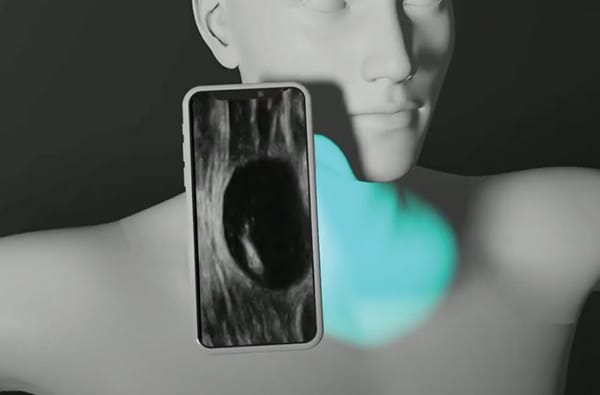
The goal is for patients to log in to QuadcordEHR via the Internet to access their pathology images and other medical images using a three-dimensional avatar of the human body or a 3D photogram of their own body. Patients’ medical images will be mapped to the avatar, and patients will be able to click the hyperlinks on the body of the avatar to pull up those images, Dr. Bui says. Users will also be able to create photograms using an app (Polycam, Altadena, Calif.) that requires them to have another person rotate a phone around their body, while they remain still, to capture all perspectives of those areas of their body that are exposed, Dr. Folio adds. Users can then zoom in on specific areas of their body to view them from an anatomical perspective. (Both technologies are featured in a YouTube video at https://youtu.be/4OjdxDGur_w.) The developers plan to offer future iterations of QuadcordEHR as a mobile phone app that would allow users to wave their phone’s camera over the area of their body for which they want to access medical images.
Mapping medical images to an avatar of a human body or a patient’s 3D photogram will help patients understand the images in an anatomical context, Dr. Bui says. For example, a patient who has had biopsies of several skin lesions would be able to click the corresponding locations on the avatar to access the pathology report for the lesions and see which ones are cancerous. This is important because the medical terminology used in pathology reports to describe locations of lesions on the body is often difficult for patients to understand, she adds.
QuadcordEHR should also help clinicians, in part by presenting a patient’s health history in an easy-to-digest format, which is particularly important when doctors see new patients, Dr. Folio says. For example, x-rays of Dr. Folio’s shoulder and ankle show osteochondritis dissecans, which resulted from him injuring bones during ski trips. But to a physician who is not familiar with Dr. Folio’s medical, occupational, and social history, these cyst-like entities in two parts of his body could appear to signal a form of bone metastasis. The ability of patients to easily access and share past x-ray images and their accompanying notes that explain the nature of these defects could prevent confusion, unwarranted testing, and patient anxiety, according to Dr. Folio.
The QuadcordEHR—pronounced “quadcorder”—takes its name from Star Trek, Dr. Folio says. The character Dr. McCoy on the long-running sci-fi television program used a medical device called a tricorder, which could sense, record, and compute medical data. Dr. Folio compares QuadcordEHR to the Star Trek tricorder, except that the real-life tool includes imaging for all image-centric medical specialties as a fourth feature. The capital letters E-H-R that compose the last syllable of the name indicate that QuadcordEHR is intended to be a type of patient-centric EHR or personal health record for images, he says.
Dr. Folio conceived the idea for QuadcordEHR when his son Lucas, an engineering student at the University of South Florida, showed him how the Polycam app can be used to scan objects and create 3D images of those objects that can be rotated on a screen. As Dr. Folio viewed the technology, he began to consider how this type of rotating 3D digital graphic could be applied to medical imaging.
Lucas Folio, who is the third partner in QuadcordEHR, created the technology’s proof of concept (or reduction to practice, in patent terms) by writing the code for QuadcordEHR in HTML5. He mapped his father’s personal medical records to the 3D avatar. The proof of concept contains not only pathology and radiology images, Dr. Folio says, but also cardiology, electrocardiogram, dermatoscopic, and intraoral images.
The three-person team applied for a patent to protect the QuadcordEHR concept and is seeking an investor to help develop a market-ready version of the product. Senior management at Moffitt has been encouraging, Dr. Folio says, and had suggested that the team seek the patent, which is pending. The developers hope the cancer center will offer the final version of QuadcordEHR to patients and that other hospitals and health systems will eventually license the technology as a tool for communicating medical information to patients.
Conceptually, QuadcordEHR aims to present medical records in the same manner that Google Earth presents neighborhood street information, Dr. Folio says. Google Earth displays a bird’s-eye view of a map and allows users to drill down to see a street or even a specific address. In a similar manner, QuadcordEHR can present a photogram of a human body that one can rotate and zoom in on and that allows patients to drill down to view, for example, a shoulder x-ray or even to drill down to the cellular level in pathology images, he explains.

Drs. Bui and Folio stress that QuadcordEHR is not only a way for patients to store medical images but a platform that physicians can use to present images to patients in a more meaningful way and that can be used in conjunction with other tools. Dr. Bui, for example, uses tools from the pathology image-sharing platform PathPresenter to enlarge digital slides and annotate them, she says. And Dr. Folio helped pioneer interactive multimedia reporting, a type of reporting used in radiology that combines images and videos with text, graphs, and hyperlinks to patient findings to improve the communication of medical information. Pathologists, radiologists, and other physicians could use these types of tools with QuadcordEHR to annotate images and add information that enhances patient understanding, he says.
As a patient-centric pathologist, Dr. Bui has seen firsthand the benefits of showing patients their medical images. In one memorable case, a patient contacted her following his third cancer diagnosis. Depressed about the finding, the patient said he wanted to see his pathology images because he needed to “stare the devil in the eyes,” she says. The experience brought home that using images to better understand disease can empower patients as well, Dr. Bui notes.
“QuadcordEHR presents an opportunity for us, as pathologists, to connect with patients,” Dr. Bui concludes. “We can connect with images, locations, and maybe educational notes that will help patients understand their diseases.”
—Renee Caruthers
Regenstrief Institute develops framework for assessing patient-matching algorithms
The Regenstrief Institute recently announced that a team of research scientists has developed an eight-point framework to assess the validity and accuracy of algorithms for linking patients’ medical records from disparate sources within and between health systems, known as patient matching.
“We recognize that the need for patient matching is not going away and that we need standardized methods to uniquely identify patients,” said Shaun Grannis, MD, MS, who led the project as Regenstrief’s vice president for data and analytics, in a press release. “Current patient-matching algorithms come in many different flavors, shapes, and sizes. To be able to compare how one performs against the other, or even to understand how they might interact together, we have to have a standard way of assessment. We have produced a novel, robust framework for consistent and reproducible evaluation. Simply put, the framework we’ve developed at Regenstrief provides a ‘measuring stick’ for the effectiveness of patient-matching tools.”
The framework’s eight-point approach, detailed in the Journal of the American Medical Informatics Association (Gupta AK, et al. 2022;29[12]:2105–2109), is a set of guidelines for creating and evaluating gold-standard data sets for measuring the performance of patient-matching algorithms. Steps one through four address recommended manual-review reporting elements for preparing a gold-standard data set and record pairs. These steps focus on data set descriptions, data preprocessing, record pair grouping, and procedures for sampling and matching record pairs. Steps five through eight address the training of human reviewers, inter-rater reliability measures, adjudication processes, and descriptions of reviewer characteristics.
“Patient identity is the last gaping hole in our health infrastructure,” said Dr. Grannis, noting that unlike many other industrialized countries, the United States does not have a national patient identifier, instead leaving the creation of unique patient identifier tools to researchers and commercial entities.
Ibex prostate cancer solution gets CE mark under IVDR
Ibex Analytics has reported that Galen Prostate is now CE-marked under the European In Vitro Diagnostic Medical Devices Regulation for supporting pathologists in the primary diagnosis of prostate biopsies.
Galen Prostate uses artificial intelligence to analyze biopsies prior to pathologist review, providing pathologists with diagnostic insights to guide diagnoses. The product’s algorithms were trained on large data sets, enriched with rare prostatic malignancies, from multiple pathology institutes worldwide.
“Galen helps pathologists diagnose cancer, provides additional insights, including a Gleason score, tumor size, and associated morphologies for each cancer slide, and offers decision-support tools to help accelerate diagnostic turnaround and reduce subjectivity,” according to a company press statement.
Galen Prostate is available for research use only in the United States.
HHS names first organizations approved to implement TEFCA
The U.S. Department of Health and Human Services has released the names of the first six organizations expected to be designated as qualified health information networks under the voluntary Trusted Exchange Framework and Common Agreement. TEFCA is an outgrowth of the 21st Century Cures Act, which required that HHS establish an infrastructure model and governing approach for nationwide health information exchange.
The organizations—Epic Systems, CommonWell Health Alliance, eHealth Exchange, Health Gorilla, Konza, and Kno2—will officially be designated as QHINS once they are fully onboarded, according to an HHS HealthITbuzz blog post.
The organizations were recognized at an HHS event last month “for their willingness to voluntarily step up and meet the rigorous TEFCA eligibility requirements, terms and conditions of TEFCA participation, and commitment to a 12-month go-live timeline,” according to the blog post. The QHIN applicants have networks that collectively cover most U.S. hospitals and tens of thousands of providers, and they process billions of transactions annually across all states, HHS reported.
The six QHINs have committed to begin exchanging data by the end of this year, Micky Tripathi, PhD, ONC national coordinator for health information technology, said at the HHS event.
The nonprofit Sequoia Project, which serves as the Office of the National Coordinator for Health Information Technology’s recognized coordinating entity for TEFCA, approved the six QHIN applications.
Dr. Aller practices clinical informatics in Southern California. He can be reached at raller@usc.edu. Dennis Winsten is founder of Dennis Winsten & Associates, Healthcare Systems Consultants. He can be reached at dwinsten.az@gmail.com.