
Editor: Frederick L. Kiechle, MD, PhD
Submit your pathology-related question for reply by appropriate medical consultants. CAP TODAY will make every effort to answer all relevant questions. However, those questions that are not of general interest may not receive a reply. For your question to be considered, you must include your name and address; this information will be omitted if your question is published in CAP TODAY.
[button size=”small” link=”http://www.captodayonline.com/q-a-submission/”]Submit a Question[/button]
Molecular testing for actionable mutations in melanoma
Written authorizations for oncologic prognostic/therapuetic tests
Molecular testing for high-grade gliomas/glioblastomas
Q. What are the actionable mutations in melanoma that warrant routine molecular testing? Who should be tested, and what specimen should be tested? Is there a role for stat BRAF testing and for next-generation sequencing?
A. Somatic activating mutations in BRAF, NRAS, or KIT are common and generally mutually exclusive in melanoma. Mutations in at least one of these genes can be found in approximately 90 percent of melanomas and each may render a patient eligible for systemic targeted therapy. BRAF- and KIT-directed agents are FDA approved. While there are no direct NRAS inhibitors, patients with NRAS-mutant melanomas may be eligible for MEK and/or PI3K inhibitors available in clinical trials, as the RAS GTPases are known to be upstream activators of both MAPK and PI3K pathways.
BRAF: Approximately 50 percent of melanomas harbor activating mutations in BRAF. Mutations at codon 600, including V600E, V600K, V600D, and V600R, are likely to respond to BRAF-targeted agents such as vemurafenib and dabrafenib. Mutations outside of codon 600 should not be treated with a BRAF inhibitor. Likewise, vemurafenib and related agents are contraindicated in patients with wild-type BRAF melanomas. Also worth noting is that BRAF mutation is not useful for diagnostic purposes because it occurs at high frequency in nevi and does not, therefore, distinguish benign melanocytic lesions from malignant.
NRAS: NRAS mutations are detectable in 15 to 25 percent of melanomas, primarily in codon 61 and less frequently in codons 12 and 13. Outside of the clinical trial setting, it is a marker we don’t know yet what to do with. Nevertheless, it is advisable to evaluate this gene because the mutation frequency is significant and therapeutic options for melanoma patients are limited.
KIT: Clinical trials demonstrate that the overall response rate to imatinib in KIT-mutated melanoma is 16 to 23 percent.1-3 These studies demonstrate the value of KIT-mutation testing in melanoma. Approximately five to 30 percent of acral and mucosal melanomas, and melanomas arising on chronically sun-damaged skin, harbor KIT mutations. Mutations occur primarily in exon 11 and less frequently in exons 13, 17, and 18.
The simple answer to the question about whom to test is any stage IV melanoma patient who is being considered for systemic therapy. At present there is little or no role for systemic targeted therapy in patients with disease limited to stages I–II (limited to primary site in skin) or III (lymph node involvement). However, clinical trials are ongoing in resected stage III disease, results of which may expand the scope of pretreatment testing to the adjuvant setting.
Regarding what specimen should be tested, in theory a metastatic tumor should be tested because this is the disease that will be treated with systemic therapy and followed for a treatment response. Indeed, there are data to suggest that testing of primary tumors and metastases may yield distinct results. Colombino M, et al.,4 showed a relatively high concordance of BRAF or NRAS mutation between primary tumor and lymph node or visceral metastasis (93 percent and 96 percent, respectively). Concordance dropped to 80 percent for brain metastasis and 75 percent for skin metastasis.
When a metastatic focus is not available for testing, the primary tumor is certainly acceptable as long as there is sufficient tumor. Because melanoma metastasizes early (Breslow depth >1 mm), it is not uncommon to see a very thin, small primary tumor submitted for testing. These early melanomas are particularly challenging to test because there is very little tumor and it is often admixed with a significant amount of inflammatory, epidermal, and dermal cells. In these cases a wild-type result may be misleading as there may be reduced sensitivity due to insufficient tumor relative to non-tumor cells. The same is true for microscopic lymph node metastases, another common specimen submitted for testing. Thus, it may be valuable to biopsy presumed metastatic foci for the purpose of confirming stage IV disease and for obtaining tumor-rich material for testing.
FNA smears may be suitable for testing as long as the test being considered is highly sensitive and appropriate for small quantities of DNA. While the nucleic acid yields are low from these slide scrapings, the DNA is often high quality because the sample was not formalin-fixed. Allele-specific PCR assays are reliable at detecting mutations in smears but are limited by the mutation-specific primers included in the assay. Approximately five to 10 percent of melanomas with activating BRAF mutations will harbor non-V600E alterations. These less common mutations could be missed in an allele-specific PCR assay that amplifies only the V600E allele. KIT testing is not appropriate for allele-specific PCR as the mutations are too variable and include indels that require alternative methods of detection. We have found that FNA smears of melanoma are amenable to amplification-based next-generation sequencing techniques. The Ion Torrent AmpliSeq system can readily amplify alleles from very small quantities of DNA such as those rendered from a smear. The advantage of testing a melanoma smear with this approach is that multiple genes can be assayed with high sensitivity from one sample. This method can detect both point mutations and small indels and, therefore, is useful for BRAF, NRAS, and KIT.
Yes, there is a role for stat BRAF testing. In fact this is a perfectly legitimate request for critical clinical scenarios. Patients with rapidly progressive disease who are highly symptomatic can benefit from prompt initiation of BRAF inhibitors when there is a detectable activating mutation. Melanoma is a cancer capable of growing rapidly in critical organs such as the brain, heart/pericardium, and GI tract (causing obstruction, bleeding). The BRAF inhibitors are capable of reducing symptoms rapidly, sometimes within days, and almost always within weeks (in responding patients). For these requests, we can process new biopsies with a stat protocol (six hours) and then triage the slides to the molecular oncology service for selection of tumor for the extraction. Our molecular laboratory prioritizes this sample and generates a pyrosequencing result by the next morning. This has been an effective strategy for preventing potentially catastrophic events such as a brain stem progression, as was imminent in one of our patients recently. Six months later his tumor was not detectable by imaging and he was symptom-free.
Similar to lung adenocarcinoma, melanoma is an ideal cancer for multigene panels. At present, some next-generation-sequencing–based oncology panels become cost-effective once three or more genes become necessary to test. Furthermore, multigene panel testing by NGS can render a timely result, such as within a 10-day turnaround time. It is quite difficult to reach that TAT with sequential, single-gene reflex testing. As with lung cancer, it is prudent to initiate systemic therapy in a stage IV melanoma patient as soon as possible, as this disease is extremely aggressive. NGS hotspot mutation panels provide the added benefit of deep sequencing, that is, detection of low frequency alleles. For KIT this is particularly important as traditional Sanger sequencing, a relatively insensitive assay, is routinely used to detect the indels that are a common mechanism of KIT mutation. NGS can also detect the more rare actionable mutations that occur in melanoma, such as PI3K and AKT, depending on what genes are included in the panel. Indeed, we recently detected a PDGFRα mutation in a tumor wild type for BRAF, NRAS, and KIT, rendering the patient eligible for imatinib or related drugs. All of these genes were sequenced in one NGS test. The PDGFRα mutation would not have been detected by the routine single-gene assays. Thus, in terms of targeted oncology panels, NGS platforms can be useful for melanoma. The role of whole genome or whole exome sequencing is not clear at this point.
- Hodi FS, Corless CL, Giobbie-Hurder A, et al. Imatinib for melanomas harboring mutationally activated or amplified KIT arising on mucosal, acral, and chronically sun-damaged skin. J Clin Oncol. 2013;31(26):3182–3190.
- Carvajal RD, Antonescu CR, Wolchok JD, et al. KIT as a therapeutic target in metastatic melanoma. JAMA. 2011;305(22):2327–2334.
- Guo J, Si L, Kong Y, et al. Phase II, open-label, single-arm trial of imatinib mesylate in patients with metastatic melanoma harboring c-Kit mutation or amplification. J Clin Oncol. 2011; 29(21):2904–2909.
- Colombino M, Capone M, Lissia A, et al. BRAF/NRAS mutation frequencies among primary tumors and metastases in patients with melanoma. J Clin Oncol. 2012;30(20):2522–2529.
Allie H. Grossmann, MD, PhD
Department of Pathology, University of Utah
ARUP Laboratories, Salt Lake City
Member, CAP Personalized Health Care Committee Work Group
Kenneth F. Grossmann, MD, PhD
Division of Oncology, Department of Medicine, Huntsman Cancer Center
University of Utah, Salt Lake City
Q. Are there generally accepted guidelines for routine molecular testing of high-grade gliomas/glioblastomas (GBM)?
A. In short, the answer is no. However, molecular testing in gliomas is on the rise in routine clinical practice, which requires the practicing pathologist to be familiar with these genetic markers, the implications of such testing, and the techniques employed for their detection.
Molecular/genetic testing typically involves answering three primary questions. Does the information help in arriving at the correct diagnosis—especially in distinguishing benign from malignant tumors? Is prognostic information gained from such testing, or can these data identify certain tumors that will be more apt to respond to specific targeted therapies or treatment strategies? In other words, can the information gleaned from molecular testing assist in analysis of tumors for diagnosis, prognosis, and/or theranostic or predictive response to current and future treatment options and modalities?
 Primary glioblastomas (1 GBM) typically arise de novo in older patients and behave aggressively. Secondary glioblastomas (2 GBM), on the other hand, develop from preceding grade II or III gliomas, typically behave less aggressively, and frequently develop in patients younger than 40 years. Recent advances in surgery, radiation therapy, and chemotherapy have provided only minor improvements in overall clinical outcomes. In time, the development of new molecular markers is expected to improve diagnostic accuracy and prognosis, as well as aid in the clinical management of GBMs.
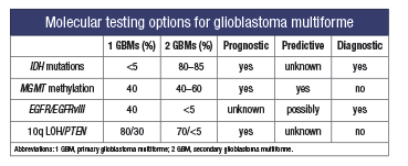
Currently, histologic evaluation remains the gold standard for glioma diagnosis; however, diagnostic difficulty may arise from tumor heterogeneity, ill-defined/overlapping morphologic features, and tumor sampling. Recently, new molecular markers have been developed, some of which have demonstrated diagnostic value, whereas others are useful as prognostic indicators for patient survival and therapeutic response. Overall, 80 percent of astrocytomas have chromosomal abnormalities. GBMs are associated with abnormalities in copy number of chromosomes 7, 9, and 10, especially gains of chromosome 7 and losses of 9p. Oligodendrogliomas are often associated with deletions of 1p and 19q, and tumors with these deletions tend to behave less aggressively and are predictive of a better response to certain chemotherapeutic options. Currently, the most common molecular testing options for high-grade gliomas include IDH1/IDH2 mutation analysis, MGMT methylation analysis, and EGFR and PTEN alteration detection analysis.
- Mutations in one of the two isozymes of isocitrate dehydrogenase (IDH) occur in approximately 40 percent of astrocytomas and oligodendrogliomas but are not seen in nonneoplastic glial tissue, and therefore can be useful in diagnosis when limited sampling is an issue. IDH mutation is associated with young age, a secondary type GBM, and increased overall survival, and it is therefore of both diagnostic and prognostic value. The mutation is generally in the IDH1 gene and is usually a point mutation in exon 4, resulting in a substitution of histidine for arginine (R132H). Sequencing can be done in formalin-fixed paraffin-embedded tissue, and there is an IHC stain for the R132H form of the protein that, when positive, can substitute for sequencing. However, 10 percent of GBMs carrying less common mutations may be missed. Real-time PCR amplification and melting curve analysis was recently reported as another method of detection using fluorescence resonance energy transfer (FRET) probes. This method is faster, less laborious, and more sensitive than sequencing.
- The MGMT gene (O6-methylguanine-DNA methyltranferase) is located at 10q26 and encodes for a DNA repair protein. Epigenetic silencing of this gene by promoter hypermethylation leads to reduced expression of the MGMT protein, which has been shown to result in improved survival in patients with GBM who are treated concurrently with the alkylating drug temozolomide and radiation therapy. The decreased MGMT protein inhibits the cells’ ability to repair alkylated DNA and thus allows alkylating drugs to work more effectively. This marker is therefore prognostic and predictive. Testing methodologies include methylation-specific polymerase chain reaction, real-time PCR, and methylation-specific pyrosequencing. Recently, the extent of MGMT methylation was proposed as a prognostic factor as well.
- EGFR (epidermal growth factor receptor) affects cell proliferation and growth. Activation of EGFR signaling through gene amplification or mutations is found in 30 to 40 percent of primary GBMs. About one-half of GBMs with EGFR amplification contain a mutant variant of the gene (EGFRvIII). Detection of either of these markers is indicative of a high-grade glioma and can be used diagnostically. The prognostic role of these markers is not currently clear. The EGFR signaling pathway is an attractive target for new chemotherapeutic agents such as anti-EGFR tyrosine kinase inhibitors. EGFR amplification can be easily detected by FISH and RT-PCR.
- Phosphatase and tensin homolog (PTEN) is a tumor-suppressor gene located on 10q23 and is frequently found in high-grade gliomas. The LOH at 10q is common in primary and secondary GBMs and anaplastic astrocytomas. PTEN mutations are found in 15 to 40 percent of primary GBMs, but they are practically absent in secondary GBMs and other gliomas. Most studies have shown 10q LOH and PTEN mutations as poor prognostic indicators for high-grade gliomas with tumor progression. LOH analysis and FISH are the methods of choice for detection.
In conclusion, some of these molecular markers can be used diagnostically to help the pathologist in glioma classification and grading, especially for tumors with ambiguous histology. Others can be used to estimate prognosis and to predict response to certain therapeutic agents. While none of these tests is ready for prime time or considered standard of care, a working knowledge of these major molecular markers and the molecular diagnostic techniques for their detection is important because their use will undoubtedly increase in routine clinical practice, especially as individualized treatment options are planned.
- Yan H, Parsons DW, Jin G, et al. IDH1 and IDH2 mutations in gliomas. N Engl J Med. 2009;360(8):765–773.
- Parsons DW, Jones S, Zhang X, et al. An integrated genomic analysis of human glioblastoma multiforme. Science. 2008;321(5897):1807–1812.
- Ichimura K, Pearson DM, Kocialkowski S, et al. IDH1 mutations are present in the majority of common adult gliomas but are rare in primary glioblastomas. Neuro Oncol. 2009;11(4):341–347.
- Weller M, Felsberg J, Hartmann C, et al. Molecular predictors of progression-free and overall survival in patients with newly diagnosed glioblastoma: a prospective translational study of the German Glioma Network. J Clin Oncol. 2009;27(34):5743–5750.
- Thompson CB. Metabolic enzymes as oncogenes or tumor suppressors. N Engl J Med. 2009;360(8):813–815.
- Everhard S, Kaloshi G, Criniere E, et al. MGMT methylation: a marker of response to temozolomide in low-grade gliomas. Ann Neurol. 2006;60(6):740–743.
- Felsberg J, Rapp M, Loeser S, et al. Prognostic significance of molecular markers and extent of resection in primary glioblastoma patients. Clin Cancer Res. 2009;15(21):6683–6693.
- Ohgaki H, Kleihues P. Genetic alterations and signaling pathways in the evolution of gliomas. Cancer Sci. 2009; 100(12):2235–2241.
- Nikiforova MN, Hamilton, RL. Molecular diagnostics of gliomas. Arch Pathol Lab Med. 2011;135(5):558–568.
- Gan HK, Kaye AH, Luwor RB. The EGFRvIII variant in glioblastoma multiforme. J Clin Neurosci. 2009;16(6):748–754.
- Mellinghoff IK, Wang MY, Vivanco I, et al. Molecular determinants of the response of glioblastomas to EGFR kinase inhibitors. N Engl J Med. 2005;353(19):2012–2024.
Mark P. Burton, MD
Medical Director, Pathology and Medical Laboratory Services
Jackson-Madison County General Hospital Medical Center Laboratory, Jackson, Tenn.
Member, CAP Personalized Health Care Committee Work Group
[hr]
Q. My hospital’s compliance consultants have advised us that pathologists may not order any oncologic prognostic/therapeutic tests that are not needed by the pathologist to establish the pathologic/cytologic diagnosis. As a result, we are requiring written authorization from one of the patient’s treating physicians before we perform testing for hormone receptors, HER2, ALK, EGFR, KRAS, BRAF, microsatellite instability, and DNA mismatch repair.
As the number of these prognostic/therapeutic tests grows, obtaining such authorizations is becoming an increasingly complex and time-consuming process that threatens to interfere with timely and efficient patient care.
Centers for Medicare and Medicaid Services regulations as published in chapter 15 of the Medicare Benefit Policy Manual say a pathologist can order only those tests that are “medically necessary so that a complete and accurate diagnosis can be reported” (80.6.5). All other tests must be ordered by a “treating physician” (80.6, 80.6.1, 80.6.2, 80.6.3, 80.6.4).
Expert guidance would be appreciated.
A. I recognize that there is debate as to whether performing hormone receptor testing is prognostic or diagnostic testing. For purposes of this question, I’ll assume that an auditor considers it to be prognostic.
One of my concerns goes beyond those definitions and is a practical one from the perspective of an attorney who deals with recoupment audits. Regardless of what any manual provision says with respect to the ability of the pathologist to order the test, if there is no documentation of the medical necessity that is convincing to the auditor, then the payment will be denied. In order for the pathologist to document medical necessity from his or her perspective for the particular patient, the medical necessity documentation almost certainly would require recitation of why the test is needed for the pathologist to complete his or her interpretive report (and this is consistent with the context of the manual provision on pathologist ordering). If the test would only be of use to the attending physician, and is not relevant to the interpretation the pathologist is providing, then the auditor could challenge the medical necessity documentation, asking how the pathologist knows that the attending physician believes the additional test would be medically necessary for the attending physician’s diagnosis and treatment of the patient (again, consistent with the context of the manual provision).
In essence, documentation of medical necessity requires that the ordering physician believes that the test/service/etc. (whatever is ordered, whether pathology, imaging, PT, etc.) is medically necessary to the ordering physician’s diagnosis and treatment of the patient. The pathologist can order a test where he or she documents “I believe this is necessary for my interpretive report for the patient.” However, the pathologist should not order a test where he or she doesn’t need the results for the interpretive report but assumes the test results would be useful for the attending physician. That decision should be made by the attending physician.
From this perspective, I believe that the hospital’s guidance and the Medicare manuals are consistent with generally recognized medical necessity principles.
Please note that there are alternatives to establishing medical necessity for the attending physician in addition to obtaining an order from the attending physician. Medical executive committee protocols and standing orders from the attending physician can also be considered.
Jane Pine Wood
Member, McDonald Hopkins LLC, Dennis, Mass.
[hr]
Dr. Kiechle is medical director of clinical pathology, Memorial Healthcare, Hollywood, Fla. Use the reader service card to submit your inquiries, or address them to Sherrie Rice, CAP TODAY, 325 Waukegan Road, Northfield, IL 60093; srice@cap.org.