
Editor: Frederick L. Kiechle, MD, PhD
Submit your pathology-related question for reply by appropriate medical consultants. CAP TODAY will make every effort to answer all relevant questions. However, those questions that are not of general interest may not receive a reply. For your question to be considered, you must include your name and address; this information will be omitted if your question is published in CAP TODAY.
[button size=”small” link=”http://www.captodayonline.com/q-a-submission/”]Submit a Question[/button]
Q. If you obtain a platelet count from a blood sample collected in a sodium citrate tube, the result is multiplied by 1.1 to correct for the volumetric difference in anticoagulant compared with EDTA. When you result the platelet count from the sodium citrate tube, is it a CAP requirement to attach a comment such as: “_#__ Results reported from blue top tube. The reference range and other method performance specifications have not been established or approved by FDA. Use results with caution.”
A. The anticoagulant of choice when testing on a hematology analyzer is EDTA (purple top). Unfortunately, EDTA can also cause in vitro platelet clumping in certain individuals. When this occurs the standard practice is to collect a specimen with sodium citrate (blue top) as the anticoagulant. The blue top specimen must be filled fully to maintain the appropriate blood-to-coagulant ratio. The platelet results are then multiplied by 1.1 to obtain the final result. It is not required to add a disclaimer since this is a mechanism to correct a problem with the specimen and the original anticoagulant.
The CAP checklist requirement follows in its entirety, including references.
There is an adequate system (such as microscopic correlation with the blood film) to prevent reporting of spurious thrombocytopenia when platelet clumps, giant platelets, or platelet satellitism are present.
NOTE: When platelet satellitosis (satellitism), significant numbers of giant platelets and/or platelet clumps are suspected/detected by cyto/histographic abnormalities or instrument rejection of a platelet result, the platelet concentration must be independently verified. Correlation with a well-prepared blood film must be made. If platelets are clumped after collection in an EDTA-anticoagulated tube that was well-mixed at the time of collection, this may represent in vitro EDTA-induced changes; platelets should be quantified from blood collected directly into a counting diluent, or by use of a different anticoagulant (e.g. liquid sodium citrate with subsequent adjustment for dilution) or by estimation from a non-anticoagulated blood film.
Evidence of Compliance:
- Written procedure defining the methods used to detect spurious thrombocytopenia or platelet abnormalities and to correct results AND
- Record showing actions taken to verify platelet concentration prior to reporting.
References
- Hyun BH, et al. Platelet satellitosis. Chicago, IL: American Society of Clinical Pathology Check Sample H-78, 1976.
- Veenhoven WA, et al. Pseudothrombocytopenia due to agglutinins. Am J Clin Pathol. 1979;72:1005-1008.
- Gloster ES, et al. Spurious platelet counts associated with bacteremia. Am J Hematol. 1985;18:329-332.
- Cunningham VL, Brandt JT. Spurious thrombocytopenia due to EDTA-independent cold-reactive agglutinins. Am J Clin Pathol. 1992;97:359-362.
- Hanseler E, et al. Estimation of the lower limits of manual and automated platelet counting. Am J Clin Pathol. 1996;105:782-787.
- Bridgen ML, Dalal BU. Cell counter-related abnormalities. Lab Med. 1999;30:325-334.
- Kunicka JE, et al. Improved platelet counting using two-dimensional laser light scatter. Am J Clin Pathol. 2000;114:283-289.
Trudy Darden,
Laboratory Accreditation Program, Technical Team Lead,
College of American Pathologists, Northfield, Ill.
[hr]
Q. Should a patient with a hematocrit greater than 55 percent be redrawn for correction always or only when prothrombin time and partial prothrombin time are elevated?
A. Accurate hemostasis and thrombosis results rely heavily on proper collection and processing (preanalytic phase) of the citrated blood specimen. Guidelines from many sources, including the Clinical and Laboratory Standards Institute,1 indicate that blood collection into nonactivating containers (e.g. polypropylene plastic or silicone-coated glass) with proper blood-to-anticoagulant ratio is required. Trisodium citrate tubes are available in a 3.2 percent or 3.8 percent concentration. A laboratory should choose a single concentration and not use the two citrate concentrations interchangeably.2 The World Health Organization and the CLSI recommend using 3.2 percent sodium citrate (105–109 nm/L), as the thromboplastin International Sensitivity Index values applied in the INR calculation are based on specimens collected in 3.2 percent citrate.2,3
The proper blood-to-anticoagulant ratio, commonly referred to as the “fill volume,” results in a 9:1 ratio of blood to anticoagulant. Underfilled tubes, defined as less than 90 percent of the fill volume, may result in prolongation of calcium-dependent, clot-based testing such as the PT and activated partial thromboplastin time (APTT) assays. Citrate’s anticoagulant effect is due to chelation of calcium in the specimen. When calcium is unavailable, coagulation cannot occur. During the analytic testing phase, a premeasured amount of calcium is added back, which allows coagulation to occur. If the citrate anticoagulant concentration is too high, the amount of calcium available during the testing phase is decreased, and the resulting time to clot formation is prolonged.
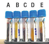
Underfilled sodium citrate tube. Citrate distributes only in plasma and if a tube is underfilled, the reduced plasma volume will contain more citrate anticoagulant, leading to increased calcium chelation (see citrate tubes A–E). Furthermore, there is an increased proportional volume of anticoagulant due to the lower plasma volume, which results in a potential dilution effect.2
The net effect is prolongation of clot-based assays (e.g. APTT, PT, protein C/protein S clot-based activity assays, and clot-based factor activity assays). The potential for erroneous results is often more pronounced in specimen tubes containing 3.8 percent sodium citrate or in smaller-volume containers such as half-draw or pediatric collection tubes.2,3
Hematocrit greater than 55 percent. Laboratories must also monitor for specimens with a hematocrit greater than 55 percent. In this situation, the plasma volume is reduced, resulting in a reduced plasma volume similar to that of an underfilled tube. The CAP hematology and coagulation checklist requires that laboratories have a written procedure for detection and special handling of specimens with elevated hematocrits.4 The amount of anticoagulant may require an adjustment in samples containing a hematocrit above 55 percent. The most efficient method to determine the appropriate citrate volume is to include a table in the laboratory specimen collection procedure indicating the modified volume of citrate for hematocrits greater than 55 percent. The formula to determine the correct volume of citrate for a given hematocrit is as follows:5
C = (1.85×10⁻³)(100−Hct)(V blood), where C indicates the volume (mL) citrate needed in the tube; Hct (%), the hematocrit of the patient; and V, the volume (mL) of whole blood. Note: In a 3.5 mL specimen tube, the volume of blood drawn is 3.15 mL (i.e. V=3.15 mL in this equation).
In theory, should an underfilled tube or specimen with a hematocrit greater than 55 percent actually show normal screening PT or APTT coagulation results, one can conclude the patient has normal hemostatic results. However, this may be a deviation from standard operating procedure, so the medical director should make the final decision. Only the laboratory medical director or a qualified laboratory physician should approve deviations from an SOP. Before a laboratory physician approves the results from an improperly filled specimen collection tube, an investigation into the patient’s clinical situation and indication for testing should be reviewed. There are situations in which re-collection may pose increased risk to a patient (a neonate, for example); in such cases performing the basic screening coagulation assays is indicated, and if the results are within normal limits, the laboratory physician may choose to release those results. Coagulation assays that do not rely on clot formation are often acceptable for testing, yet approval for testing should also be determined by the pathologist.
Other calcium-dependent, clot-based special coagulation assays such as the protein C (PC) and protein S (PS) activity assays, which use PT or APTT methodology, should not be performed on an over-citrated specimen. In the PC and PS assays, falsely prolonged clotting times may result in overestimation of PC or PS activity. Thus, an over-citrated specimen secondary to an underfilled tube or hematocrit greater than 55 percent may be misinterpreted as having normal PC or PS activity. In the clot-based PC and PS activity assays, a short time to the clotting endpoint (fibrin formation) correlates to abnormally low PS and/or PS activity. Therefore, low PC and/or PS activity results from specimens that are underfilled or have hematocrit greater than 55 percent may represent a true, “qualitatively” low value, yet the accuracy (i.e. exact value) of the activity would be in question; if testing is performed, the pathologists must review these results and determine how (or if) the result should be reported. Clot-based factor activity assays are also subject to these preanalytic errors. Over-citrated specimens may cause falsely prolonged clotting times in the factor activity assays, resulting in erroneously low factor activity levels.
Overfilled sodium citrate tube. Overfilled specimen collection tubes can also occur. A common cause for overfilled specimen tubes is when whole blood is first drawn into a syringe and then forcibly injected into a specimen tube. Overfilled citrate tubes can also occur when the stopper (cap) is removed and additional blood is added. This may be an indication that a specimen from one collection tube was poured (added) into a second collection tube, and this practice is unacceptable.2 Overfilled tubes may result in poor mixing of the anticoagulant and lead to inaccurate results. Regardless of the cause of an overfilled tube, it violates the 9:1 ratio, and testing should be approved only by the pathologist.
In conclusion, improperly filled specimen tubes should call into question the collection process. The guidelines provided by CLSI H21-A5 clearly state, “Specimens that are clotted, collected in the wrong anticoagulant type (e.g., EDTA, sodium heparin), have other than a 9:1 blood to anticoagulation ratio, or are collected or stored in a container with an activating surface, are not suitable for testing and should be rejected [italics added].”1 One must be cautious when accepting an improperly collected specimen. Approving the specimen for testing often undermines the strict specimen collection requirements designed for accurate coagulation test results and patient safety. Making exceptions to these rules may falsely reassure clinical medical providers that improperly filled citrate specimen tubes are acceptable. Exceptions to the re-collection for underfilled tubes or patients with hematocrit greater than 55 percent should be considered only when screening PT and/or when APTT test results are normal or when specimen re-collection is not possible.
- Clinical and Laboratory Standards Institute. Collection, Transport, and Processing of Blood Specimens for Testing Plasma-Based Coagulation Assays and Molecular Hemostasis Assays; Approved Guideline—Fifth Edition (H21-A5). Jan. 23, 2008.
- Kitchen S, Olson JD, Preston FE, eds. Quality in Laboratory Hemostasis and Thrombosis. 2nd ed. Chichester, West Sussex, U.K.: Wiley-Blackwell; 2013:45–56.
- Bennett ST, Lehman CM, Rodgers GM, eds. Laboratory Hemostasis: A Practical Guide for Pathologists. 2nd ed. Cham, Switzerland: Springer International; 2015: 173–175.
- College of American Pathologists. Hematology and coagulation checklist. July 28, 2015.
- Kottke-Marchant K. An Algorithmic Approach to Hemostasis Testing. 2nd ed. Northfield, Ill.: CAP Press; 2016:46–47.
Andrew Jackson Goodwin IV, MD, Associate Professor, Department of Pathology, University of Vermont,
College of Medicine, Burlington, Member, CAP Coagulation, Resource Committee
[hr]
Dr. Kiechle is medical director of clinical pathology, Memorial Healthcare, Hollywood, Fla. Use the reader service card to submit your inquiries, or address them to Sherrie Rice, CAP TODAY, 325 Waukegan Road, Northfield, IL 60093; srice@cap.org. Those questions that are of general interest will be answered.