Reading COVID-19’s signature: lung tissue injury
Anne Paxton
July 2020—Alain Charles Borczuk, MD, began his practice of pathology as a resident 28 years ago and has spent quite a bit of his career doing autopsies. But it is this year, during the pandemic, that he’s finding some of the best applications of his autopsy work as he seeks to understand the lung injury patterns in SARS CoV-2, or COVID-19 patients.
“There has not been a period of time in which there’s been a greater realization of the absolute critical importance of doing autopsy than right now,” says Dr. Borczuk, professor of pathology at Weill Cornell Medical College and vice chair of anatomic pathology at Weill Cornell Medicine.
One reason: “We’re not really getting tissue-based samples in the majority of COVID-19 patients during their clinical course, and autopsy gives us a way to learn about the disease that we haven’t had before. Everyone has looked at these autopsies and said, ‘In looking at these patterns, in talking about what we are seeing, it has resonated with the things we’re finding clinically.’”

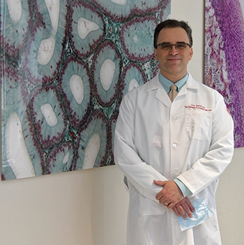
Dr. Alain Charles Borczuk: “The question I am interested in,” he says, “is what is the unique injurious effect on the lung that is resulting in this quite accelerated severe disease with thrombosis.”
When asked to discuss COVID-19 and lung tissue injury, Dr. Borczuk cautions that his viewpoint is narrow. From the autopsy suite, his observations relate to COVID patients who don’t recover from their illness. “It’s a bit of a skewed perspective,” he admits. “We are seeing treatment failures, not treatment successes.” And as he notes, the majority of hospital admissions for COVID lead to discharge, not death.
The percentage of COVID patients who are intubated is also a figure outside his knowledge. “I find those numbers to be complicated ones that, as pathologists, we don’t get to learn. We have an idea of how many patients are in intensive care, we know total patients on ventilators, but we don’t actually know how dynamic that is—how many people are coming off the ventilators or never getting on the ventilators to begin with.”
“The death rate associated with COVID is a highly controversial number because of the different populations and how much testing is done. So it’s difficult to tell, from the perspective of a pathologist, exactly what those numbers are.”
Nevertheless, Dr. Borczuk’s experience based on patient mortality from COVID offers useful insights into one of the most mystifying elements of COVID: Exactly what does the disease do to the patient’s lungs? In his view, it’s a question that can be fully answered only with an autopsy.
The decline in the number of autopsies performed in recent years, he says, is partly owing to the improvements in clinical diagnostic testing: imaging, serum testing, noninvasive testing, even biopsies, which are done more easily now than ever before.
“We’re able to know a lot more about what is going on in a patient without doing an autopsy. As a result, there is a feeling among the people who consent for autopsy and the clinicians who know the patients and the patients’ families that they know everything that happened already and there’s no point to an autopsy. And I’ve heard that said.”
Still, “autopsies often reveal severity of disease that was unexpected, and other things that were never detected, often to the surprise of the clinicians when they get the information. But that fact alone hasn’t increased the number of autopsies.”
With COVID-19, he says, “There is so much uncertainty around this disease. It’s new and for the folks who are sick it’s a very severe disease. Families are left with a situation where they have a lot of unanswered questions, and so do the physicians. And that combination has led to interest in doing autopsy.”
“I can tell you what we find in patients who are acutely ill, who are given respiratory support in a variety of ways including ventilation, but who progress and who pass away.” It’s an older population and they tend to have comorbid illnesses, which vary quite a bit but include a history of hypertension, of diabetes, of hyperlipidemia, says Dr. Borczuk, who spoke with CAP TODAY on May 1.
Among those patients, “we are seeing a variety of lung injuries. There is often inflammation and even ulcerating lesions in the upper airway, trachea, and bronchus. The patients who have been on ventilators have more of the changes in the lung that we might expect with organization of the lung and recovery steps that are involved in lung healing, meaning the person was supported past that acute period into a more chronic or subacute period, so the lung has a chance to regenerate.”
The phase of acute respiratory distress syndrome that he sees is organizing diffuse alveolar damage. “So we see ongoing injury alongside the healing.” But he cannot be sure a particular injury might be from the ventilator. “It could be that they’ve been supported by a ventilator and now have time to heal and then have a new injury on top of that healing. Or it may well be an effect of viral infection. We just don’t know.”
The injury that is resonating with the clinicians who have taken care of the patient is a lesion of the large airways that is inflammatory, he says. ”The clinicians describe a lot of mucus plugging. We haven’t seen as much of that, but we certainly have seen a lot of large-airway epithelia injury with some ulceration and inflammation, so it would not surprise me if there is also a lot of mucus production. We just haven’t seen a lot of it at autopsy. It may be because they are taking care of the mucus plugging clinically, so it is less dramatic at the time of autopsy.”
 CAP TODAY Pathology/Laboratory Medicine/Laboratory Management
