Anne Paxton
February 2013—If anybody is a believer in programs to reduce hemolysis rates in the hospital, it’s Dennis Ernst, MT(ASCP), director of the Center for Phlebotomy Education. Ever since he left the bench 15 years ago, Ernst has been traveling the country with a mission: to show clinical laboratories, nursing departments, hospital administrators, and clinicians that the payoff from high-quality phlebotomy is much greater than they might realize. Despite hemolysis being the No. 1 reason the laboratory rejects blood specimens, hemolysis does not strike randomly, and it’s not inevitable, Ernst emphasizes. “Typically the causes of hemolysis are all behavioral,” he says.
But even Ernst was taken aback at the dazzling results of a Lean process improvement program launched to tackle hemolysis rates about four years ago at Sarasota (Fla.) Memorial Health Care System. As powerful as Lean principles have proved to be, it isn’t often that they produce seven-figure savings to an institution’s bottom line. “At Sarasota, they decided hemolysis was not only a threat to patient quality of care, but a threat to the facility’s well-being. They rolled up their sleeves and said ‘we’re not going to take it anymore.’ And it’s incredible what they projected their savings to be when they really got serious,” Ernst says.
It all started with the laboratory confronting a stark fact: Blood specimens collected in Sarasota’s Emergency Care Center (ECC) and throughout the 806-bed hospital had significantly higher hemolysis rates than those collected by phlebotomy staff. In a 2009 count, “the hemolysis rate facility-wide was about three percent, while the ECC had a rate of about eight percent,” says Charlene Harris, FACHE, MT(ASCP), director of laboratory services. When hemolysis was measured by nursing unit, only two of 18 units were meeting the goal of keeping hemolysis at two percent or below.

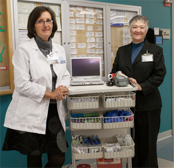
Dana Rickard (left) and Charlene Harris at Sarasota Memorial, where a phlebotomist was assigned to collect blood in sections of the ECC for a period, and the hemolysis rate dropped. “That’s what convinced them that it really made a difference,” Harris says. [Photo: Kenneth Gronquist]
At Sarasota, the laboratory decided to conduct a positive improvement study, says Dana J. Rickard, MT(ASCP), preanalytical manager. In the summer of 2009, with the help of Charlotte Damato, a Six Sigma/Lean Quality coach and expert in work and role redesign, “we did a value stream analysis of turnaround time in the lab in connection with the ECC.” In addition, Harris says, “we did some Lean studies of physical workflow in the ECC and the lab to see structurally where improvements could be made. So we used Lean and everything that goes along with that methodology.”
Representatives from Becton Dickinson, which provides the hospital’s blood collection supplies, worked with the process improvement team to arrive at a non-confrontational way to observe how blood was being drawn, Rickard says. “The BD consultants didn’t directly watch; they would individually ask the nurses, technologists, and phlebotomists to explain the blood collection supplies they would use and how they would collect the specimen. So it wasn’t as though the laboratory was actually coming to nursing to ask how blood was being drawn. And that helped tremendously.”
BD consultants determined through observation that staff were using several different procedures to collect blood, but most blood specimens were collected using an 18- to 20-gauge IV catheter. To collect the blood from the catheter, most staff attached a multi-sample adapter and Single Use Holder. In some cases, blood was collected off the catheter using a syringe, then transferred into tubes using a straight needle. For repeat tests, sometimes specimens were collected by flushing the catheter and then drawing blood from the catheter.
But an early meeting of BD consultants, catheterization lab staff, and nursing staff showed that the nurses believed that after they drew the blood, the laboratory was doing something that caused the red cells to burst, Rickard says. So the challenge to the laboratory was clear. “We had to figure out a way to get the nursing staff on board with how the specimens should be drawn.”
“Hemolysis is probably one of the greatest preanalytical errors that occurs to specimens that arrive at the laboratory,” says Frederick L. Kiechle, MD, PhD, medical director of clinical pathology at Memorial Healthcare System, Hollywood, Fla., and editor of the CAP’s phlebotomy guide, So You’re Going To Collect a Blood Specimen. Among other problems, the concentration of potassium inside red blood cells is much higher than in plasma, and ruptured red cells add potassium to the specimen. It’s important that potassium be normal before surgery, so one danger is that hemolysis may mislead clinicians to rely on falsely high potassium levels in laboratory results.The earlier trend toward decentralization of phlebotomy in many hospitals—the model employed at Sarasota—has not generally been good for controlling hemolysis, Dr. Kiechle believes. “Phlebotomy is no longer being done by a team that resides under the control of the laboratory. Nurses and sometimes doctors on all the floors have this function, and the level of training varies a lot.”
While some facilities can make decentralized phlebotomy work, Ernst agrees it’s exceedingly difficult. “In larger facilities, you’ll see an attempt to decentralize phlebotomy as a way to make the staff more efficient, when in fact that’s really not what the outcome is. Plenty of studies have shown that in general it’s a failed concept. By and large, most facilities don’t have the time or resources to implement or maintain it successfully.” That’s one reason why, at least for blood cultures, the Centers for Disease Control and Prevention recommends that a dedicated team of phlebotomists be responsible for performing draws.
Theoretically, any sample could have a slight degree of hemolysis. “It has to be pretty severe before you even see the change in the color of the serum, because even unhemolyzed serum can be a shade of yellow that borders on pink, so it’s hard to visually determine,” Ernst says. Many analyzers now chemically determine how much hemoglobin is in the serum, so if the hemoglobin exceeds a certain threshold, personnel can be notified and make a judgment on whether or not the test result is being influenced. On the other hand, if the sample is tested as whole blood and not centrifuged, “then the hemolysis would not even be detected and the test results could be reported without any knowledge that the sample is hemolyzed.”
Just how easily red blood cells can hemolyze is one of the major teaching points Dr. Kiechle emphasizes when trying to educate people about phlebotomy. “For someone who has perhaps not been doing phlebotomy for a while or has never done it, it’s very important to realize that just having patients clench their fist a number of times, or keeping pressure on the patient’s arm with a tourniquet for too long, can lead to rupture of these red cells.”
If people think of red blood cells as fragile crystal orbs, they might have an appreciation for how delicate phlebotomy needs to be to avoid hemolysis, Ernst says. “Once you respect the fragility of red blood cells, you’re naturally more careful with them. You don’t pull really hard on a plunger syringe, you don’t draw from IV devices if you can help it, and you don’t force blood into a tube when you’re emptying a syringe into it.”
Exposure to alcohol, perhaps still left on the skin before the draw is performed, is one chemical problem that can cause blood specimens to hemolyze. But many cases of hemolysis can be explained in terms of fluid dynamics and the mechanical effects of temperature, shear rate, or pressure. Shear forces, created by any flow of the specimen fluid, generate friction. The more force exerted to move the fluid, the more shear it encounters, and the more likely red blood cells, which are deformable particulates in the fluid, will be ruptured.
Sometimes these forces can hemolyze samples in ways that aren’t all that obvious. For example, in breaking the seal on a syringe, in the manufacturing process the manufacturer has “seated” the plunger, Ernst explains. “So that plunger is kind of stuck on arrival. If you don’t ‘unseat’ the plunger and it’s still somewhat adhering to the barrel of the syringe, and you draw the sample, then the unseating of the plunger causes an instant increase in the force with which the blood is pulled. There is this rapid negative pressure on the first mL of blood in the sample that’s drawn, so the cells rush into the syringe rather than being gently coaxed.”
A sluggish draw, occurring when the needle is not centered in the vein, generally means the bevel opening of the needle is partly in the vein and partly in the vein walls. Or, as also frequently happens, a vein may collapse onto the needle. “Therefore, the narrow opening the red cells have to pass through is partially occluded, and you have the same effect as if you were drawing through a very narrow cannula,” Ernst says. “Whenever the blood just trickles, then you know that something is occluding the needle and probably hemolyzing the sample.”
The addition of air to a sample can also cause hemolysis, Ernst says. “When the device used to draw the blood is not properly fitted—for example, the syringe is not properly fitted to the vascular access device—then air gets into the fitting, it foams up the blood, and instead of having nothing but straight liquid blood in your syringe, you end up with a bubbly mixture.” Hemolysis can occur because this “frothing” introduces more turbulence, or shear, into the sample as it’s withdrawn.
At times, phlebotomists “milk” capillaries without realizing that squeezing the skin, too, can cause hemolysis. “When phlebotomists are performing a capillary puncture or finger stick and didn’t pre-warm the site, they may attempt to excessively squeeze the site or milk blood out of the puncture, and whenever you’re doing that, you’re forcing red cells through the incision. Any kind of force is unfriendly to red cells, and that includes forcefully milking blood out of the site.”
 Shaking the tubes after they are filled with specimen is another way to expose them to excessive pressure and cause hemolysis. For related reasons, Ernst has found, sometimes the culprit may be the design of a pneumatic tube system used to transport samples. He was asked to visit a health care facility in the Southwest where the tube manufacturer was being blamed for hemolyzed samples. “We came in and did a thorough audit of what the samples were being subjected to, and we eliminated everything except the pneumatic tube system. We watched the phlebotomists draw blood on morning rounds, and we drew two samples of the same type of tube but walked one sample down and pneumatically transported the other one.” That’s how they ascertained that the pneumatically transported samples were more susceptible to hemolysis, and they devised a measure to fix the problem. “We found the pneumatic tube system had really dramatic drops and turns, so we recommended they add better padding to the canisters.”
 CAP TODAY Pathology/Laboratory Medicine/Laboratory Management
