 CAP TODAY Pathology/Laboratory Medicine/Laboratory Management
June 2023—Three initiatives are underway at the American Board of Pathology, one of which its CEO Gary Procop, MD, MS, describes as “a new era of transparency and collaboration.”
Read More »Home >> Tag Archives: Cytopathology/histology (see also Cytopathology in Focus)
Against all odds, a histology lab in rural Ghana
February 2023—I was fortunate last summer to be part of a team established to build a histology laboratory in rural Ghana. Despite our many challenges, we succeeded in building a laboratory where specimens can now be processed for pathological interpretation. Although many obstacles remain, this laboratory offers great potential to improve the pathology services provided to a large portion of rural Ghana.
Read More »Cytopathology in focus: Protocol for reporting cervicovaginal cytology specimens
August 2022—The protocol for the reporting of cervicovaginal cytology, the first in a series of CAP cytopathology protocols, became available for use in a synoptic format on June 22. This protocol is a collaborative effort, based on input from past and present members of the CAP Cytopathology Committee and prepared in conjunction with the CAP Pathology Electronic Reporting Committee. It was presented via webinar to the CAP House of Delegates on March 31. A two-week open comment period followed; all comments were reviewed and appropriate changes were incorporated into the protocol.
Read More »Fluid cytology—key features and ancillary testing
June 2022—What to look for in serous fluid cytology is what Eva M. Wojcik, MD, of Loyola University in Chicago, and Xiaoyin “Sara” Jiang, MD, of Duke Health, set forth in their CAP21 session last year.
Read More »Cytopathology in focus: Special stains in the cytology laboratory
January 2022—A consistent virtue of the cytopathology laboratory is that it combines two qualities essential to patient care: It provides an accurate and timely diagnosis. The ability to make a prompt diagnosis is particularly important in immunosuppressed or otherwise vulnerable patient populations for whom a timely diagnosis can result in early treatment initiation and potentially better outcomes.
Read More »Cytopathology in focus: p16 immunostaining in cytology specimens—a diagnostic pitfall
January 2022—Cytopathologists are often the first pathologists to diagnose HPV-related head and neck squamous cell carcinomas (HNSCC). These head and neck cancers can present as superficial masses amenable to fine-needle aspiration, where p16 immunostaining is used as a surrogate marker for HPV in situ hybridization in a subset of squamous cell carcinomas.
Read More »Cytopathology in focus: Statistical reporting—benefits beyond the numbers
January 2022—The CAP has a robust Laboratory Accreditation Program with a commitment to continually improving the programs and providing appropriate resources needed for compliance. As a deemed status organization, validation surveys are performed annually through the Centers for Medicare and Medicaid Services and the feedback obtained provides direction for education.
Read More »Cytopathology in focus: Our appeal to program participants—return glass slides
January 2022—A diverse and copious inventory of Pap and nongynecologic glass slides is the backbone of the CAP glass slide educational programs. Each year tens of thousands of cytopathology slides are packaged and mailed to laboratories enrolled in CAP educational glass slide programs throughout the world. Prior to mailing, numerous cytotechnologists and cytopathologists screen these slides and companion web enhancement images to ensure quality and diagnostic accuracy.
Read More »Cytopathology in focus: Virtual education in cytology: pandemic silver lining
January 2021—Although the challenges we face due to the COVID-19 pandemic are significant, adapting to our new circumstances can be a driver for positive change. Cytology education had started using virtual learning resources in the past few years with great variation among institutions and countries. The pandemic forced the community to embrace virtual learning in a variety of modalities. These changes may lead to lasting improvements in the quality and accessibility of educational opportunities for cytology trainees, and not just in the United States. In the same way, we are witnessing the exponential growth of digital pathology as applied to surgical pathology. In cytopathology the applications have been limited to telecytopathology in the setting of rapid on-site evaluation (ROSE) for interventional procedures. The difficulties in dealing with multilayered focus have been given as reasons not to pursue whole slide imaging for cytology. As the cytology community has, even if reluctantly, gained experience with remote sign-out, we may see a push for digital imaging for primary diagnosis in cytology.
Read More »Cytopathology in focus: Telecytology for rapid on-site evaluation
January 2021—Rapid on-site evaluation (ROSE) for cytology specimens is performed at many institutions to improve the quality of health care by proper triage of obtained material to increase the diagnostic yield, or to direct appropriate investigation. It also helps to control health care costs by reducing the rate of nondiagnostic specimens, unnecessary passes, and repeat procedures. The number of procedures requiring ROSE is growing due to the increase in the number of platforms used to perform minimally invasive procedures.
Read More »Cytology workload limits: For adequacy assessments, it’s time, not slides

August 2018—The CAP and the Centers for Medicare and Medicaid Services reached an understanding earlier this year on how adequacy assessments and rapid on-site evaluations in cytology can be accounted for without causing undue impact on workload limits. The agreement, communicated to state survey agency directors in a March 16 CMS memorandum, is reflected in the updated CAP accreditation program cytopathology checklist released this month.
Read More »In free CytoAtlas app, 750 images for 100+ diagnoses

October 2014—Like many cytopathology trainees, Charanjeet Singh, MD, who recently completed a cytopathology fellowship at MD Anderson Cancer Center in Houston, found it challenging at times to find classic examples of entities to learn from and to study for exams. Most texts he consulted contained just one or two images of a particular diagnosis. And the material in training programs from all specialties varies. Even though there is a large volume of cytology cases at MD Anderson, for example, it wasn’t enough to learn gynecologic cytology, which is why he pursued an elective rotation at Houston Methodist Hospital.
Read More »New programs next year in gyn, nongyn cytopathology
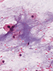
November 2013—Participants in the CAP cytopathology programs will have new modules to select and new cases to learn from in 2014. Samples of static images that accompany the Touch Imprint/Crush Prep cases. In gynecologic cytopathology, a new L module for education will feature liquid-based SurePath and ThinPrep slide methods only. These will be designated PAPL/APAPL, with a choice of series one or two.
Read More »